Sunday, May 28, 2023

Altered 5-HT2A/C receptor binding in the medulla oblongata in the sudden infant death syndrome

Haynes RL, Trachtenberg F, Darnall R, Haas EA, Goldstein RD, Mena OJ, Krous HF, Kinney HC. Altered 5-HT2A/C receptor binding in the medulla oblongata in the sudden infant death syndrome (SIDS): Part I. Tissue-based evidence for serotonin receptor signaling abnormalities in cardiorespiratory- and arousal-related circuits. J Neuropathol Exp Neurol. 2023 May 25;82(6):467-482. doi: 10.1093/jnen/nlad030. PMID: 37226597; PMCID: PMC10209647.

Abstract

The sudden infant death syndrome (SIDS), the leading cause of postneonatal infant mortality in the United States, is typically associated with a sleep period. Previously, we showed evidence of serotonergic abnormalities in the medulla (e.g. altered serotonin (5-HT)1A receptor binding), in SIDS cases. In rodents, 5-HT2A/C receptor signaling contributes to arousal and autoresuscitation, protecting brain oxygen status during sleep. Nonetheless, the role of 5-HT2A/C receptors in the pathophysiology of SIDS is unclear. We hypothesize that in SIDS, 5-HT2A/C receptor binding is altered in medullary nuclei that are key for arousal and autoresuscitation. Here, we report altered 5-HT2A/C binding in several key medullary nuclei in SIDS cases (n = 58) compared to controls (n = 12). In some nuclei the reduced 5-HT2A/C and 5-HT1A binding overlapped, suggesting abnormal 5-HT receptor interactions. The data presented here (Part 1) suggest that a subset of SIDS is due in part to abnormal 5-HT2A/C and 5-HT1A signaling across multiple medullary nuclei vital for arousal and autoresuscitation. In Part II to follow, we highlight 8 medullary subnetworks with altered 5-HT receptor binding in SIDS. We propose the existence of an integrative brainstem network that fails to facilitate arousal and/or autoresuscitation in SIDS cases.

Tuesday, May 23, 2023

BRAF mutations

Kenney-Jung DL, Rogers DJ, Kroening SJ, Zatkalik AL, Whitmarsh AE, Roberts AE, Zenker M, Gambardella ML, Contaldo I, Leoni C, Onesimo R, Zampino G, Tartaglia M, Battaglia DI, Pierpont EI. Infantile epileptic spasms syndrome in children with cardiofaciocutanous syndrome: Clinical presentation and associations with genotype. Am J Med Genet C Semin Med Genet. 2022 Dec;190(4):501-509. doi: 10.1002/ajmg.c.32022. Epub 2022 Nov 29. PMID: 36448195; PMCID: PMC9825647.

Abstract

Gene variants that dysregulate signaling through the RAS-MAPK pathway cause cardiofaciocutaneous syndrome (CFCS), a rare multi-system disorder. Infantile epileptic spasms syndrome (IESS) and other forms of epilepsy are among the most serious complications. To investigate clinical presentation, treatment outcomes, and genotype-phenotype associations in CFCS patients with IESS, molecular genetics and clinical neurological history were reviewed across two large clinical research cohorts (n = 180). IESS presented in 18/180 (10%) cases, including 16 patients with BRAF variants and 2 with MAP2K1 variants. Among IESS patients with BRAF variants, 16/16 (100%) had sequence changes affecting the protein kinase domain (exons 11-16), although only 57% of total BRAF variants occurred in this domain. Clinical onset of spasms occurred at a median age of 5.4 months (range: 1-24 months). Among 13/18 patients whose IESS resolved with anti-seizure medications, 10 were treated with ACTH and/or vigabatrin. A substantial majority of CFCS patients with IESS subsequently developed other epilepsy types (16/18; 89%). In terms of neurodevelopmental outcomes, gross motor function and verbal communication were more limited in patients with a history of IESS compared to those without IESS. These findings can inform clinical neurological care guidelines for CFCS and development of relevant pre-clinical models for severe epilepsy phenotypes.

Xie MG, Wang XF, Qiao J, Zhou J, Guan YG, Li TF, Qi XL, Luan GM. The clinicopathological features of ganglioglioma with CD34 expression and BRAF mutation in patients with epilepsy. Front Mol Neurosci. 2023 Feb 23;16:1022364. doi: 10.3389/fnmol.2023.1022364. PMID: 36910263; PMCID: PMC9995901.

Abstract

Objective: The aim of the study was to evaluate the clinicopathological features, as well as the surgical prognosis, of epilepsy-associated gangliogliomas (GG) with CD34 expression and BRAFV600E mutation.

Methods: Clinical data of patients who underwent epilepsy surgery for GG were retrospectively studied. Univariate and multivariate analyses were performed to evaluate the correlations of clinical and pathological factors with molecular markers of CD34 expression and BRAFV600E mutation in GG.

Results: A total of 208 patients with GG had immunohistochemical detection of CD34 expression (positive/negative: 184/24), and among them, 89 patients had immunohistochemical detection of BRAFV600E mutation (positive/negative: 54/35). By univariate and multivariate analyses, seizure aura (p = 0.025), concordance of ictal electroencephalogram (EEG) findings (p = 0.045) and medial temporal tumor (p = 0.030) were found to be related to CD34 expression, but only hospitalization time (p = 0.042) was different for BRAF-mutated status. In addition, drug-resistant epilepsy (p = 0.040) and concordance of interictal EEG findings (p = 0.009) were found to be associated with tumor progression-free survival (PFS) in univariate analysis, but only concordance of interictal EEG findings was with significance in multivariate analysis. However, CD34 expression or BRAFV600E mutation in GG was not found to be associated with surgical outcomes of seizure control and tumor PFS.

Conclusion: The CD34 expression or BRAFV600E mutation in GG may partly influence the distribution of clinicopathological features of patients with epilepsy, but they may be not able to predict the surgical prognosis of seizure outcome and tumor recurrence.

Xie M, Wang X, Qiao J, Zhou J, Guan Y, Li T, Qi X, Luan G. The clinical and pathological features of low-grade epilepsy-associated glioneuronal tumors. Sci Rep. 2022 Oct 28;12(1):18163. doi: 10.1038/s41598-022-22443-2. PMID: 36307486; PMCID: PMC9616895.

Abstract

The aim of the study was to evaluate the clinicopathological features, as well as the surgical prognosis, of epilepsy-associated glioneuronal tumors (GNT) with CD34 expression and BRAF mutation. Clinical data of patients who underwent epilepsy surgery for GNT were retrospectively studied. Univariate and multivariate analyses were performed to evaluate the correlations of clinical and pathological factors with molecular markers of CD34 expression and BRAFV600E mutation in GNT. A total of 247 patients with GNT had immunohistochemical detection of CD34 expression (CD34 positive vs. negative: 198/49), and among them, 102 patients had immunohistochemical detection of BRAFV600E mutation (BRAF positive vs. negative: 59/43). Univariate analysis found that tumor types (P < 0.001), patient population (P = 0.015), seizure aura (P = 0.007), drug-resistant epilepsy (P = 0.036), concordance of ictal electroencephalogram (EEG) findings (P = 0.032), surgical resection extent (P = 0.045), tumor location (P = 0.007) and duration of epilepsy (P = 0.027) were related to CD34 expression, and that concordance of ictal EEG findings (P = 0.031) and age at surgery (P = 0.015) were related to BRAFV600E mutation. In addition, history of generalized tonic-clonic seizure (HR 0.12; P = 0.035), drug-resistant epilepsy (HR 0.13; P = 0.030) and concordance of interictal EEG findings (HR 8.01; P = 0.039) were associated with tumor progression-free survival (PFS). However, CD34 expression or BRAFV600E mutation in GNT was not associated with surgical outcomes of seizure control and tumor PFS. The CD34 expression or BRAFV600E mutation in GNT may partly influence the distribution of clinicopathological features of patients with epilepsy, but they may be not able to predict the surgical prognosis of seizure outcome and tumor recurrence.

Sunday, May 21, 2023

Frontotemporal dementia

Iowa woman, 27, has 99% chance of developing dementia: ‘I needed to know’

Alyssa Nash inherited the gene mutation for FTD, a rare form of early-onset dementia — but here's why she has hope for her future

Alyssa Nash, 27, of Northern Iowa, is a healthy, energetic young woman with a bright smile and a deep love for her family. She enjoys ceramics, painting and working as a receptionist at a local hospital.

But underneath her happy exterior, Nash carries a quiet fear of turning into someone she no longer recognizes.

Seven years ago, when she was just 20, Nash learned she's a carrier for a gene mutation that can cause FTD (frontotemporal dementia), an early-onset neurodegenerative disease that most commonly impacts people as early as their 30s, 40s and 50s.

FTD is marked by the degeneration of the frontal and/or temporal lobes of the brain, according to the Association for Frontotemporal Degeneration (AFTD).

Much less common than Alzheimer’s disease, FTD currently affects some 60,000 people in the U.S.

Symptoms can start as early as 21 years old, but most cases occur between the ages of 45 and 64, per the AFTD.

Common signals of the disease include personality changes, difficulty speaking and understanding language, trouble with decision-making and a lack of empathy.

Dr. Tiffany Chow, neurologist and vice president of clinical development at the biotechnology company Alector in South San Francisco, California, is an expert in this type of dementia.

"Unlike Alzheimer's disease, FTD causes a drastic turnaround."

"FTD is one of the most fascinating neurodegenerative diseases because it actually affects your personality," she told Fox News Digital in an interview. 

"Unlike Alzheimer's disease, FTD causes a drastic turnaround," Chow said. "So the nicest people become the most obnoxious people. It's a marked difference that affects everything in terms of how the world perceives you and how you perceive the world."

From a clinical standpoint, FTD is devastating to families, the doctor said.

"These are people who are mainly in their late 40s or 50s," she said. "They're still working and may be very active in their community. They're still raising children. And so in some ways, the impact is much greater economically and in terms of the ripple effects on the people around them."

It’s a challenge that Nash knows all too well. Her father began to display signs of FTD when he was only 38 years old, just as Nash was entering high school.

A long road to diagnosis

"The biggest challenge was seeing him change completely into someone else and become someone I never thought he could be," Nash told Fox News Digital in an interview. "And having to grow up in that atmosphere — it was tough."

It was seven years before Nash’s father was officially diagnosed. 

"We went through years of unknowns and questions," Nash said.

Along with her mother and brother, Nash helped care for her dad as his dementia progressed, even as she struggled to hide his condition from everyone at her high school.

"It was this big secret I was keeping," she said. "I basically had two lives. I didn’t tell anyone at school because I didn't want to be different — I wanted to feel normal, in a sense."

She added, "But then I'd have to go home right away to help take care of my dad and make sure he was OK."

Delayed diagnoses are common

The majority of dementia cases — more than 60%, studies show — are not diagnosed for a host of reasons, according to Diane Ty, senior director of the Milken Institute Center for the Future of Aging in Washington, D.C.

And by the time they are identified, the condition has usually progressed to the later stages, she said.

Ty had a similar experience when her own father began exhibiting symptoms of dementia. 

"We should be doing cognitive testing as part of primary care."

"We confused some of the behavioral challenges that we were seeing as the effects of him taking early retirement and being depressed, but in retrospect, it was the early stages of dementia," she said during a discussion with Fox News Digital.

In most cases, Ty said, it’s not until there is some sort of accident or incident that the affected person is brought in for testing.

"That’s just not acceptable," she said. "We should be doing cognitive testing as part of primary care, not unlike getting a mammogram or a colonoscopy. It should be part of the annual wellness visit."

Ty added, "We are not regularly looking at our brain health. I think that's one of the biggest barriers to timely detection and diagnosis."

Genetic screening: ‘I needed to know’

In some cases, FTD can be passed down from a parent to a child. 

"We have made a lot of progress in determining the genetic mutations that seem to be responsible for frontotemporal dementia, more so than in Alzheimer's disease," Chow explained. 

Most genetic FTD cases are caused by a mutation in one of three genes: C9ORF72, MAPT or GRN.

Chow noted that 30% to 40% of patients living with FTD carry these genetic mutations or variants. 

"That's where genetic counseling becomes important," she said. "The genetic counselor will often talk about the pros and cons of knowing this information and run through the scenarios if the test comes out positive."

"When they said I had the option for genetic testing, I said, ‘Sign me up.’"

As soon as Nash’s father was diagnosed and Nash found out the condition was genetic, she knew she wanted to find out whether she was a carrier of the mutation.

"I needed to know, because for so many years, I wasn't in control," she told Fox News Digital. "I needed to make a plan. This was a chance to be in control of myself."

She added, "When they said I had the option for genetic testing, I said, ‘Sign me up.’"

Tough news leads to empowerment

When Nash found out in 2016 that she's a carrier of the gene mutation, she had mixed emotions.

On one hand, she was scared of what that might mean for her future. 

"I did take it really hard when I first found out," she said. "It took years before I could speak about it publicly."

At first, Nash had processed the news as if it was a diagnosis. But over the years, she found that the knowledge helped her appreciate even the smallest details of day-to-day life.

"Now I feel empowered by this knowledge," Nash told Fox News Digital. "It's going to help more than I could possibly imagine. That flip of the brain was definitely needed."

Nash said she appreciates having the ability to share her discovery with her loved ones — the people who would be most affected, ultimately, if her predisposition led to a diagnosis down the road.

Nash said she is hopeful that dementia treatments may become available in time to benefit her.

"I wouldn’t want my worst enemy to have to go through what I went through without having the answers," she said.

Dementia treatments in the pipeline

Although there are therapies that can treat the symptoms of dementia — in an attempt to make people less anxious or less restless — there are no disease-modifying therapies currently approved by the FDA, Chow said.

"There are several experimental therapies being studied that are designed to target specific genetic mutations for FTD," said Chow.

Alector, the biotechnology company where Chow works in drug development, is currently running advanced clinical trials of a medication called latozinemab.

"Although it’s still under investigation, the drug may boost aspects of our system to clear the abnormal protein collections [in the brain] that are believed to cause FTD," Chow said.

Given her young age, Nash said she is hopeful that dementia treatments may become available in time to benefit her.

"That time difference presents an opportunity to apply a disease-modifying therapy once one has been approved, because the earlier you start a therapy that has been proven efficacious and safe, the better chance you have of not developing symptoms once you reach 50 or 60," Chow said. 

"And that’s what makes it so empowering."

Ty is also optimistic about the potential for dementia treatments — not only by discovering new drugs, but also repurposing existing medications.

"In the next 10 years, I hope to see exciting changes in the way we detect, diagnose and treat dementia."

"A recent survey showed that there's a reluctance among physicians to have the conversation and do the testing because there’s this assumption that ‘there’s nothing we can do,’" she said. "But I think that's going to change with the introduction of some of these new therapeutics."

"In the next 10 years, I hope to see exciting changes in the way we detect, diagnose and treat dementia," she added.

There are also ways to delay dementia development through lifestyle modifications, Ty pointed out.

A 2020 study published in The Lancet identified 12 modifiable risk factors for dementia, including smoking, physical inactivity, social isolation, depression, air pollution, lack of education, hearing loss, traumatic brain injury, hypertension, high alcohol consumption, diabetes and obesity.

"If those were managed, you could reduce 40% of dementia worldwide," Ty said.

Looking ahead: ‘Excited for my future’

For those who are facing a similar struggle, Nash’s advice is to not bottle up all the emotions that come with it and to seek mental health care.

"Put your feelings out there," she recommended. "It's okay not to be okay at times. It’s a heavy thing to carry, but knowledge is power."

"You can make a plan," she went on. 

"For me, I’m going to do everything I can to help people and to help myself."

For a long time after discovering her mutation, Nash had dwelled on the idea that she "only had so many years" before her dementia symptoms would begin. Over the past couple of years, however — with the help of her family support system and regular counseling sessions — she has adopted a new mindset.

"I found myself again," she said. "I have a gosh-darn future, and I can’t wait for it."

https://www.foxnews.com/health/iowa-woman-27-99-percent-chance-developing-dementia-needed-know

Friday, May 19, 2023

Cytokines in new-onset refractory status epilepticus predict outcomes

Article In Brief

Patients with new-onset refractory status epilepticus had significantly higher serum levels of several key pro-inflammatory cytokines than study participants with other forms of refractory status epilepticus, suggesting that immune therapies targeting specific aspects of the innate immune system may be helpful for treating patients with cryptogenic NORSE.

Innate immunity-related inflammation plays a key role in the pathogenesis and outcomes of new-onset refractory status epilepticus (NORSE), according to a new international multicenter study led by investigators from Yale University School of Medicine published in Annals of Neurology in March.

NORSE is a rare, confounding clinical entity with a high mortality rate of 16 to 27 percent in adults and around 12 percent in children, the study authors pointed out. It strikes suddenly and without warning, often in young, previously healthy individuals. In the initial presentation, patients experience a sudden onset of continuous seizures or a flurry of very frequent seizures, without any apparent cause, that do not respond to standard anticonvulsant medications. Typically, they require prolonged anesthesia with coma-inducing drugs to get the seizures under control.

The majority of patients who survive usually live with major long-term neurocognitive and functional disabilities, often including drug-resistant epilepsy. About half of all patients with NORSE have an unknown etiology for their condition even after extensive workups, referred to as cryptogenic NORSE.

Study Details

In the new study, patients with NORSE had significantly higher serum levels of several key pro-inflammatory cytokines compared with study participants with other forms of refractory status epilepticus (RSE). The findings suggest that immune therapies targeting specific aspects of the innate immune system, such as IL-6 blockers or anti-CXCL8, may be helpful for treating patients with cryptogenic NORSE.

The study enrolled 61 patients—15 of whom were cryptogenic—at 13 hospitals in the United States, one in Canada, and one in France, all between February 2013 and July 2022. The patients with NORSE were at least 2 years old. The NORSE patient cohort included 24 patients with a prior febrile illness between one and 14 days prior to the onset of RSE, a subtype of NORSE known as febrile infection-related epilepsy syndrome (FIRES). Investigators also enrolled 37 patients with other forms of RSE from known etiology, 12 patients with autoimmune encephalitis without status epilepticus, 22 patients with drug-resistant epilepsy who had a seizure (but not SE) within 24 hours before sample collection, and 18 control patients without epilepsy.

The researchers collected serum samples for all patients and cerebrospinal fluid samples from 29 patients with NORSE, 14 with RSE, 10 with autoimmune encephalitis, and 17 controls.

Pro-Inflammatory Cytokines

The serum analysis showed overproduction of pro-inflammatory cytokines/chemokines in the serum and in the CSF of patients with status epilepticus compared with patients without status epilepticus. They found differences among the five subgroups of patients for nine serum cytokines/chemokines and five CSF cytokines/chemokines.

Patients with NORSE had significantly higher serum levels of CXCL8, MIP-1alpha, and CCL2 and significantly higher CSF levels of IL-1beta than those with other forms of RSE. Those with cNORSE also had significantly higher serum levels of CXCL8, MIP-1alpha, and CCL2 compared to all non-cryptogenic RSE.

https://journals.lww.com/neurotodayonline/Fulltext/2023/05180/The_Role_of_Inflammation_in_New_onset_Refractory.2.aspx

Hanin A, Cespedes J, Dorgham K, Pulluru Y, Gopaul M, Gorochov G, Hafler DA, Navarro V, Gaspard N, Hirsch LJ. Cytokines in New-Onset Refractory Status Epilepticus Predict Outcomes. Ann Neurol. 2023 Mar 5. doi: 10.1002/ana.26627. Epub ahead of print. PMID: 36871188.

Abstract

Objective: The objective of this study was to investigate inflammation using cerebrospinal fluid (CSF) and serum cytokines/chemokines in patients with new-onset refractory status epilepticus (NORSE) to better understand the pathophysiology of NORSE and its consequences.

Methods: Patients with NORSE (n = 61, including n = 51 cryptogenic), including its subtype with prior fever known as febrile infection-related epilepsy syndrome (FIRES), were compared with patients with other refractory status epilepticus (RSE; n = 37), and control patients without SE (n = 52). We measured 12 cytokines/chemokines in serum or CSF samples using multiplexed fluorescent bead-based immunoassay detection. Cytokine levels were compared between patients with and without SE, and between the 51 patients with cryptogenic NORSE (cNORSE) and the 47 patients with a known-etiology RSE (NORSE n = 10, other RSE n = 37), and correlated with outcomes.

Results: A significant increase of IL-6, TNF-α, CXCL8/IL-8, CCL2, MIP-1α, and IL-12p70 pro-inflammatory cytokines/chemokines was observed in patients with SE compared with patients without SE, in serum and CSF. Serum innate immunity pro-inflammatory cytokines/chemokines (CXCL8, CCL2, and MIP-1α) were significantly higher in patients with cNORSE compared to non-cryptogenic RSE. Patients with NORSE with elevated innate immunity serum and CSF cytokine/chemokine levels had worse outcomes at discharge and at several months after the SE ended.

Interpretation: We identified significant differences in innate immunity serum and CSF cytokine/chemokine profiles between patients with cNORSE and non-cryptogenic RSE. The elevation of innate immunity pro-inflammatory cytokines in patients with NORSE correlated with worse short- and long-term outcomes. These findings highlight the involvement of innate immunity-related inflammation, including peripherally, and possibly of neutrophil-related immunity in cNORSE pathogenesis and suggest the importance of utilizing specific anti-inflammatory interventions

Wednesday, May 17, 2023

Could this patient have Aicardi-Goutieres syndrome type 7?

A 5 1/2 year old boy at 3 1/2 years began losing lower extremity strength, initially on the right but then bilaterally. Initially, there also seemed to be involvement of right upper extremity and right sided hyperreflexia. He gradually lost his ability to ambulate independently.  There was also loss of verbalization. Currently, the deep tendon reflexes are brisk diffusely and motor tone is increased diffusely. 

He had an autism/ID Xpanded panel done through GeneDx.  This showed a variant of unknown significance in the LAS1L gene. He also had a cerebral palsy spectrum disorder panel of genetic testing done, which showed a variant of unknown significance in the IFIH1 gene [c.2644A>C (p.Met882Leu)] This variant had been detected on an earlier autism/ID panel, which was performed as a trio of tests with both of his parents. His father had the same variant, causing the autism/ID panel not to report it on the initial findings.  Whole genome sequencing was nondiagnostic. Some of the previously identified VUS were re-demonstrated. Cerebrospinal fluid studies were also unremarkable, except for an increased CSF neopterin of 123 (normal 7 to 65).  A repeat neopterin was 43 (7-40). Extensive additional laboratory studies were unrevealing.

The IFIH1 gene is associated with Aicardi-Goutieres syndrome type 7. There has been speculation that this, indeed, is the disorder the patient has.

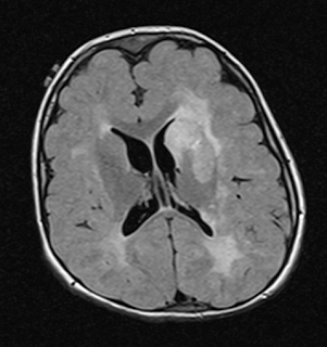
Images from the most recent brain MRI are below. The axial and coronal are T2 and the sagittal T1. Aside from the thinning of the posterior corpus callosum, no abnormality was described.


Uggetti C, La Piana R, Orcesi S, Egitto MG, Crow YJ, Fazzi E. Aicardi-Goutieres syndrome: neuroradiologic findings and follow-up. AJNR Am J Neuroradiol. 2009 Nov;30(10):1971-6. doi: 10.3174/ajnr.A1694. Epub 2009 Jul 23. PMID: 19628626; PMCID: PMC7051307.

Abstract

Background and purpose: To date, few studies have focused specifically on imaging findings in Aicardi-Goutières syndrome (AGS). We set out to evaluate retrospectively neuroradiologic data from a large sample of patients with AGS, focusing on the pattern of white matter abnormalities and the temporal evolution of the cerebral involvement to establish the radiologic natural history of the disease.

Materials and methods: Thirty-six patients, 18 girls and 18 boys, were included. All had a clinical diagnosis of AGS, genetically confirmed in 31 of them. For every subject, we reviewed at least 1 CT and 1 MR imaging study; 19 (52.7%) had multiple examinations. In all, we reviewed 109 examinations. Clinical-neuroradiologic comparisons were analyzed by using the chi(2) test.

Results: Calcifications were found in all subjects, mainly in the basal ganglia, lobar white matter, and dentate nuclei. Abnormal white matter was present in all the subjects, showing 2 patterns of distribution: diffuse in 18 (50%) and an anteroposterior gradient in 18 (50%). Cystic areas were observed in the temporal and/or frontal lobes in 12/36 patients (33.3%). A correlation was found between early age at onset and severity of the leukoencephalopathy in the frontal (P = .024) and temporal (P = .034) regions. A significant degree of cerebral atrophy was found in 31/36 subjects (86.1%). The neuroradiologic presentation remained substantially stable with time.

Conclusions: The different neuroradiologic presentations of AGS are here outlined for the first time in a large sample of patients. These findings may facilitate more precise and earlier diagnosis of this rare but probably underdiagnosed syndrome.

Han VX, Mohammad SS, Jones HF, Bandodkar S, Crow YJ, Dale RC; AGS-JAKi Study Group. Cerebrospinal fluid neopterin as a biomarker of treatment response to Janus kinase inhibition in Aicardi-Goutières syndrome. Dev Med Child Neurol. 2022 Feb;64(2):266-271. doi: 10.1111/dmcn.15025. Epub 2021 Aug 20. PMID: 34415581.

Abstract

Janus kinase (JAK) 1 inhibition represents a precision medicine approach in the treatment of Aicardi-Goutières syndrome (AGS), through targeting of type I interferon-mediated cell signalling. Blood interferon mRNAseq has been proposed as a biomarker of disease with utility in therapeutic monitoring. Objective cerebrospinal fluid (CSF) biomarkers tracking treatment efficacy are currently lacking. Here, we report a retrospective case series of 13 patients (median age 6y, range 2y 6mo-17y; five females, eight males) with AGS demonstrating significantly elevated CSF neopterin levels at first sampling (median 200nmol/L, range 45-2024nmol/L), compared to 13 age-matched controls with non-inflammatory neurological conditions (median 23nmol/L, range 5-34nmol/L, p<0.001). Five patients with AGS treated with JAK inhibitors demonstrated a median 81.5% reduction of CSF neopterin (range -36% to -88% change from baseline), compared to eight untreated patients with AGS demonstrating a median 7% reduction in CSF neopterin (range -63% to +117% change) (p=0.047). Our data indicate a biological effect of JAK inhibitors, and the potential role of CSF neopterin as a biomarker of treatment response.

The latter citation was previously entered as https://childnervoussystem.blogspot.com/2021/03/janus-kinase-inhibition-in-aicardi.html

Any correspondence to gbreningstall@gillettechildrens,com

Tuesday, May 16, 2023

Mom was tough

From a childhood friend of mine

After my mom died in November of 2013, we held a wonderful celebration at St. John's Cathedral. She is interred in the 'Walk of Souls' there. Students from mom's first years as a teacher at Merrill Jr. High, some in their eighties, made sure to attend, as well as students from South High, where she taught after she left Hill.

Yes, my mom was a great teacher, engaging, funny, smart, memorable. She had enough love for all her students, but still was a great mom to Pam and me.

But, you have no idea how strong and brave my mom was...

In the mid-50's, mom had to commit her mother to the state hospital in Pueblo. Mom was recently divorced. Grandma was living with us, but it was even obvious to this little guy that grandma was not well. We didn't have much money, so that was the only option. Mom drove to Pueblo at least once a month to visit. Pam and I were never allowed to see grandma. We were told grandma had 'Huntingtons Chorea', a degenerative disease of the brain stem.

Some years later, mom was taking classes at DU to get her Master's in education. During a lecture, the professor was talking about hereditary diseases. Huntington's was mentioned. If your parent has it, you have a 50-50 genetic chance. After the lecture, mom ran down to tell the professor that her mom had it and wanted to ask more questions. He didn't look at her, slammed his notebook shut and walked off. (I remember his name to this day, the coward). Mom later spoke of him with a snarl.

So, here is a divorced mother with two kids, and she has a chance to get a most cruel disease.

What to do? When you have a twitch in bed, is that it? When you spill a glass, do you have it? If you have it, then your kids also have a 50-50 chance. I can't imagine the years of worry that mom endured, though she only spoke of it once it became clear that she was perhaps free of the scourge when she was in her mid-40's. It meant that my sister and I dodged a hideous fate.

Mom found a nice couple who would take Pam and me if she got the sickness. They lived in the Sloan's Lake area.

Imagine arranging to have your children taken away.

Mom was tough.

Efficacy and safety of dietary therapies for childhood drug-resistant epilepsy

Devi N, Madaan P, Kandoth N, Bansal D, Sahu JK. Efficacy and Safety of Dietary Therapies for Childhood Drug-Resistant Epilepsy: A Systematic Review and Network Meta-analysis. JAMA Pediatr. 2023 Mar 1;177(3):258-266. doi: 10.1001/jamapediatrics.2022.5648. PMID: 36716045; PMCID: PMC9887534.

Abstract

Importance: Despite advances in the understanding of dietary therapies in children with drug-resistant epilepsy, no quantitative comparison exists between different dietary interventions.

Objective: To evaluate the comparative efficacy and safety of various dietary therapies in childhood drug-resistant epilepsy.

Data sources: Systematic review and network meta-analysis (frequentist) of studies in PubMed, Embase, Cochrane, and Ovid published from inception to April 2022 using the search terms ketogenic diet, medium chain triglyceride diet, modified Atkins diet, low glycemic index therapy, and refractory epilepsy.

Study selection: Randomized clinical trials comparing different dietary therapies (ketogenic diet, modified Atkins diet, and low glycemic index therapy) with each other or care as usual in childhood drug-resistant epilepsy were included. Abstract, title, and full text were screened independently by 2 reviewers.

Data extraction and synthesis: Data extraction was conducted following Preferred Reporting Items for Systematic Reviews and Meta-analyses reporting guideline. Cochrane risk-of-bias tool was used to assess the study quality. Effect sizes were calculated as odds ratio with 95% CI using random-effects model. The hierarchy of competing interventions was defined using the surface under the cumulative ranking curve.

Main outcomes and measures: Short-term (≤3 months) 50% or higher and 90% or higher reduction in seizure frequency and treatment withdrawal due to adverse events were the primary efficacy and safety outcomes.

Results: Of 2158 citations, 12 randomized clinical trials (907 patients) qualified for inclusion. In the short term, all dietary interventions were more efficacious than care as usual for 50% or higher seizure reduction (low glycemic index therapy: odds ratio [OR], 24.7 [95% CI, 5.3-115.4]; modified Atkins diet: OR, 11.3 [95% CI, 5.1-25.1]; ketogenic diet: OR, 8.6 [95% CI, 3.7-20.0]), while ketogenic diet (OR, 6.5 [95% CI, 2.3-18.0]) and modified Atkins diet (OR, 5.1 [95% CI, 2.2-12.0]) were better than care as usual for seizure reduction of 90% or higher. However, adverse event-related discontinuation rates were significantly higher for ketogenic diet (OR, 8.6 [95% CI, 1.8-40.6]) and modified Atkins diet (OR, 6.5 [95% CI, 1.4-31.2]) compared with care as usual. Indirectly, there was no significant difference between dietary therapies in efficacy and safety outcomes.

Conclusions and relevance: This study found that all dietary therapies are effective in the short term. However, modified Atkins diet had better tolerability, higher probability for 50% or higher seizure reduction, and comparable probability for 90% or higher seizure reduction and may be a sounder option than ketogenic diet. Direct head-to-head comparison studies are needed to confirm these findings.

Activity and recovery among youth with concussion: A meta-analysis

Rochelle Chauhan, Anika Cheng, Rebecca Tsow, Brodie M. Sakakibara, Shelina Babul, Julia Schmidt. Activity and Recovery Among Youth With Concussion: A Meta-analysis. Pediatrics (2023) 151 (5): e2022059592. https://doi.org/10.1542/peds.2022-059592

CONTEXT

Concussions in youth are highly prevalent. Previously, rest was prescribed to prevent adverse outcomes; however, early return to activity is emerging to improve the recovery trajectory.

OBJECTIVES

To evaluate the effectiveness of early return to physical and social activity interventions on recovery outcomes in youth with concussion.

DATA SOURCES

A systematic review was conducted up to October 2022.

STUDY SELECTION

We included randomized controlled trials (RCTs) and non-RCTs that reported effects of activity-based interventions on symptoms, quality-of-life (QoL), and return to preinjury activity levels in children and youth after a concussion.

DATA EXTRACTION

Three authors independently extracted data on publication year and country, study setting and design, sample size, participant demographics, intervention, outcome(s), and author conclusion. Meta-analysis was conducted on appropriate RCTs.

RESULTS

Twenty-four studies were included in the final review, of which 10 were RCTs. There was a significant effect of activity interventions on symptom reporting (standardized mean difference, 0.39 [95% confidence interval, 0.15 to 0.63]; I2, 0%; P = .002). There was not a significant effect of activity-based interventions on QoL (mean difference, −0.91 [95% confidence interval, −7.76 to 5.94]; I2, 0%; P = .79). No meta-analysis was performed on return to preinjury activity levels because of insufficient number of RCTs conducted.

LIMITATIONS

One outcome was excluded from the meta-analysis. Interventions emphasizing social activity were lacking.

CONCLUSIONS

Findings indicate that activity-based interventions may significantly improve concussion symptoms. There is insufficient data to understand the effect of activity-based intervention on QoL and return to preinjury activity levels.

Thursday, May 11, 2023

Neuroradiological findings in children with subdural hematoma and suspected abusive head trauma

Zahl, S.M., Andersson, J., Wester, K. and Wikström, J. (2023), Neuroradiological findings in children with subdural hematoma and suspected abusive head trauma. Ann Child Neurol Soc, 1: 44-52. https://doi.org/10.1002/cns3.3

Abstract

Aim

Abusive head trauma (AHT) is often suspected in infants with subdural hematoma (SDH). Other neuroradiological findings have also been reported in assumed AHT, such as hypoxic–ischemic injury (HII), cortical vein thrombosis, and subarachnoid hemorrhage. The purpose of this study was to investigate neuroradiological and clinical findings in cases of suspected AHT.
Methods

Infants with SDH suspected to be caused by AHT, referred to the Swedish National Board of Forensic Medicine during the period 1994–2018, were considered for inclusion.
Results

Ninety-six cases were included, with 68% males. The proportions of infants born prematurely, and twins, were higher than in the normal population. Signs of “benign enlargement of the subarachnoid space” (BESS) were found in 36% and were associated with chronic SDH and nonacute symptoms. HII was found in 16% and was associated with subarachnoid hemorrhage and high mortality, but not traumatic findings. Clinical signs of increased intracranial pressure were associated with retinal hemorrhages.
Conclusion

This analysis indicates that infants with SDH investigated for AHT are a heterogeneous group with some cases associated with external signs of trauma and others with possibly nontraumatic etiologies. Infants with BESS are more likely to have chronic SDH and nonacute symptoms. Patients with HII often have serious/fatal outcomes, but are not associated with external signs of injury. The overrepresentation of males, premature infants, and twins is unexplained.

From the article:

Pre-existing BESS/chronic SDH

As recently shown in Andersson et al., a study on the same population, infants with chronic SDH had a male overrepresentation and were in comparison to acute SDH children more often premature, had lower mortality, and had more often increased head circumference. Andersson et al. also showed that there were clinical and/or radiological signs of BESS as a possible cause of the hemorrhage in a substantial portion of these children. None of the acute SDH infants had any sign of BESS. In addition, our results show that BESS is associated with nonacute symptoms, while skull fractures are associated with acute SDH. These findings further support the assumption launched by Andersson et al. of acute SDH and chronic SDH as two fundamentally different conditions...

Conclusion

In infants with SDH and suspected AHT, we found different groups of neuroradiological, epidemiological, and clinical findings, suggesting different etiologies.

Some infants have an obvious traumatic etiology; with acute SDH, skull fracture(s) or other signs of trauma, and AHT should then be suspected if a credible history of accidental trauma is not presented.

In some infants with chronic SDH, underlying BESS seems to represent a possible cause for the SDH.

Infants with hypoxic–ischemic injury seldom have external signs of trauma and have an even sex distribution. The overall high proportion of boys and premature infants may be related to different vulnerability factors and warrants further investigation, as also goes for the pathophysiology of hypoxic–ischemic injury in these infants.

Wednesday, May 10, 2023

What is this?


8/1/13 


8/28/15 


                                                                              4/11/19


6/25/20


                                                                          9/16/20


                                                                           1/22/21


                                                                          12/13/22


10 year old female (4/29/13) with intractable epilepsy and profound motor and cognitive handicap. Has had extensive and unrevealing laboratory investigations. Two brain biopsies performed.  In 01/2021, she had a Stealth-guided stereotactic brain biopsy through a left frontal bur hole.  The biopsy showed white matter tracts with scattered calcifications.  The Ki-67 proliferative index was slightly elevated.  "These findings are not conclusive and we cannot rule out the possibility of neoplastic process."  She then had a repeat brain biopsy on 04/21/2021.  There were dystrophic calcifications in the subcortical white matter and extensive reactive gliosis seen involving the basal ganglia.  This was a biopsy of the left caudate.  There was no evidence of a neoplastic process.  The report indicates, "However, the findings on clinical examination and imaging studies are highly suggestive of an unusual neurocutaneous syndrome, particularly hypomelanosis of Ito." The patient does have cutaneous findings akin to those of hypomelanosis of Ito.

Any thoughts can be conveyed to gbreningstall@gillettechildrens.com.


Cluster headache and migraine have circadian features

Cluster headaches and migraines were associated with the circadian system, according to a recent systematic review and meta-analysis in the journal Neurology.

“These results raise the potential for using circadian-based treatments for headache disorders,” said study author Mark Joseph Burish, MD, PhD, University of Texas Health Science Center at Houston in a news release. “This could include both treatments based on the circadian rhythm—such as taking medications at certain times of the day—and treatments that cause circadian changes, which certain medications can do.”

Researchers sourced studies from medical databases including Medline Ovid, Embase, PsycINFO, Web of Science, and Cochrane Library, then 2 researchers independently conducted the systematic review/meta-analysis while following the Preferred Reporting Items for Systematic Reviews and Meta-Analysis (PRISMA) guidelines. A separate genetic analysis was conducted to study the circadian pattern of expression in Clock Controlled Genes (CCGs) by cross-referencing genome-wide association studies (GWAS) of headache, a non-human primate study of CCGs in a variety of tissues, and recent reviews of brain areas relevant in headache disorders.

By combining the results of both studies, researchers were able to catalog the circadian features at different levels, including behavioral, systems, and cellular levels, providing a holistic understanding of the role of CCGs in headache disorders.

The meta-analysis found 1513 studies, 72 of which met inclusion criteria, while the genetic analysis found 16 GWAS, 1 non-human primate study, and 16 imaging reviews. Findings revealed significant differences between the circadian patterns of attacks in cluster headache and migraine.

In cluster headaches, a circadian peak was observed between 21:00 and 03:00, while in migraine, a circadian trough was observed between 23:00 and 07:00. The circannual peaks for cluster headache were observed in spring and fall, while migraine peaks were observed between April and October. The chronotype was highly variable across studies for both types of headaches. At the systems level, lower melatonin and higher cortisol levels were reported for cluster headache, while lower urinary melatonin levels were observed in migraine—even lower during an attack. At the cellular level, cluster headache was associated with core circadian genes CLOCK and REV-ERBα, and 5 of the 9 cluster headache susceptibility genes were CCGs. Conversely, migraine was associated with core circadian genes CK1δ and RORα, and 110 of the 168 migraine susceptibility genes were CCGs.

“This reinforces the importance of the hypothalamus—the area of the brain that houses the primary biological clock—and its role in cluster headache and migraine,” Dr Burish concluded. “It also raises the question of the genetics of triggers such as sleep changes that are known triggers for migraine and are cues for the body’s circadian rhythm.”

Authors noted that they did not have information on factors that may influence the circadian cycle, such as comorbid disorders or medications, which may have limited study findings.

https://www.hmpgloballearningnetwork.com/site/neuro/news/migraines-cluster-headaches-associated-circadian-system

Benkli B, Kim SY, Koike N, Han C, Tran C, Silva E, Yan Y, Yagita K, Chen Z, Yoo SH, Burish MJ. Circadian Features of Cluster Headache and Migraine: A Systematic Review, Meta-analysis, and Genetic Analysis. Neurology. 2023 Mar 29:10.1212/WNL.0000000000207240. doi: 10.1212/WNL.0000000000207240. Epub ahead of print. PMID: 36990725.

Abstract

Background and objectives: Cluster headache and migraine have circadian features at multiple levels (cellular, systems, and behavioral). A thorough understanding of their circadian features informs their pathophysiologies.

Methods: A librarian created search criteria in Medline Ovid, Embase, PsycINFO, Web of Science, and Cochrane Library. Two physicians independently performed the remainder of the systematic review/meta-analysis using PRISMA guidelines. Separate from the systematic review/meta-analysis we performed a genetic analysis for genes with a circadian pattern of expression (Clock Controlled Genes or CCGs) by cross-referencing genome-wide association studies (GWAS) of headache, a non-human primate study of CCGs in a variety of tissues, and recent reviews of brain areas relevant in headache disorders. Altogether, this allowed us to catalog circadian features at the behavioral level (circadian timing, time of day, time of year, and chronotype), systems level (relevant brain areas where CCGs are active, melatonin and corticosteroid levels), and cellular level (core circadian genes and CCGs).

Results: For the systematic review and meta-analysis, 1513 studies were found and 72 met inclusion criteria; for the genetic analysis we found 16 GWAS, 1 non-human primate study, and 16 imaging reviews.

Cluster headache: Behaviorally, meta-analyses showed a circadian pattern of attacks in 70.5% (3490/4953) of participants across 16 studies, with a clear circadian peak between 21:00-03:00 and circannual peaks in spring and autumn. Chronotype was highly variable across studies. At the systems level, lower melatonin and higher cortisol levels were reported. At the cellular level, cluster headache was associated with core circadian genes CLOCK and REV-ERBα, and five of the nine cluster headache susceptibility genes were CCGs.

Migraine: Behaviorally, meta-analyses showed a circadian pattern of attacks in 50.1% (2698/5385) of participants across eight studies, with a clear circadian trough between 23:00-07:00 and a broad circannual peak between April-October. Chronotype was highly variable across studies. At the systems level, urinary melatonin levels were lower in migraine participants and even lower during an attack. At the cellular level, migraine was associated with core circadian genes CK1δ and RORα, and 110 of the 168 migraine susceptibility genes were CCGs.

Discussion: Cluster headache and migraine are highly circadian at multiple levels, reinforcing the importance of the hypothalamus. This review provides a pathophysiological foundation for circadian-targeted research into these disorders.

Friday, May 5, 2023

Infantile spasms: A frontline guide for pediatricians

Eric H Kossoff, Seva G. Khambadkone. Infantile spasms: A frontline guide for pediatricians
Contemporary PEDS JournalApril 2023

From the article

Home video recording as a clinical tool

Given the length of a typical clinic visit, it is unlikely that the infant will have a spasm in the office. This is where home video recordings (eg, via smartphones) can be extremely helpful. Home video recording, first advised by the Child Neurology Society to streamline IESS management at the onset of the COVID-19 pandemic, has since been endorsed as a continued recommendation toward timely intervention. In preparing to evaluate a patient with possible IESS, pediatricians should ask caregivers to record suspected events. Videos should be reviewed prior to the scheduled visit if possible and filed to share with consulting providers including child neurologists. Shortened clips can easily be shared through patient/provider portals in most electronic medical records, whereas longer videos may require secure cloud services...

Follow-up and prognosis

Unfortunately, even in some cases with spasm resolution, long-term prognosis is often poor. Outcomes include subsequent epilepsy and neurodevelopmental impairment. Etiology may be the most important predictor of outcome.23 Lead time to treatment is another critical and modifiable prognostic factor. Several studies to date have found improved developmental/intellectual outcomes with shorter delays between spasm onset and treatment, highlighting the importance of early intervention. In the UKISS study, this relationship was suggested to be dose dependent, where longer lead time durations were associated with a stepwise decline in developmental assessment scores at aged 4 years.

Children with history of IESS should be followed closely for developmental and neurological sequelae. Psychomotor development should be monitored until at least kindergarten, even in children who appear to have excellent prognoses. It is best to involve a developmental pediatrician in this care as well as to have a low bar for the recruitment of allied specialists including physical, occupational, and speech therapists. While outcomes can be challenging, close allyship among the family, primary pediatrician, neurologist, and broader care team will go far toward creating an environment of support and resilience.

https://www.contemporarypediatrics.com/view/infantile-spasms-a-frontline-guide-for-pediatricians

Wednesday, May 3, 2023

The interaction between ketogenic diet therapy and anti-seizure medications

Armeno ML, Kossoff EH. Let food be thy medicine. The interaction between ketogenic diet therapy and anti-seizure medications: A systematic review. Epileptic Disord. 2023 Mar 28. doi: 10.1002/epd2.20055. Epub ahead of print. PMID: 36987562.

Abstract

Ketogenic diet therapy (KDT) is a nonpharmacological treatment that has been demonstrated to be effective in reducing seizures in patients with drug-resistant epilepsy. As the majority of patients on KDT are also receiving anti-seizure medications (ASMs), questions about their combination often arise. KDT is typically implemented as an add-on, and not a substitute for ASMs. Drug monitoring and specific laboratory studies may be helpful in specific cases of cotherapy. Valproate, topiramate, zonisamide, and lamotrigine may be potentially problematic with KDT, but the evidence for this is not conclusive. ASM reduction is usually attempted after 1 month of KDT if a child is showing seizure reduction (but weaning ASMs does not require seizure freedom). Failure to wean an ASM does not mean KDT has failed and adding a new ASM may be beneficial in those cases after several months of KDT fine-tuning. The purpose of this review was to discuss the evidence for possible negative (or positive) pharmacodynamic interactions between KDT and ASMs. In addition, practical suggestions for the weaning or adding of ASMs in patients on KDT are provided.

Autism spectrum disorder prevalence rises among minority children

The rate of children diagnosed with autism spectrum disorder (ASD) continues to increase, particularly in non-white children, a CDC surveillance summary finds, highlighting the need for enhanced infrastructure that provides equitable diagnostic, treatment, and support services.

“For the first time among children aged 8 years, the prevalence of ASD was lower among White children than among other racial and ethnic groups, reversing the direction of racial and ethnic differences in ASD prevalence observed in the past,” authors wrote in the report. “Black children with ASD were still more likely than White children with ASD to have a co-occurring intellectual disability.

The Autism and Developmental Disabilities Monitoring (ADDM) Network is a surveillance program consisting of 11 sites across the United States that estimates the prevalence of ASD among children aged 8 years. Staff review developmental evaluations and records from community medical and educational service providers to formulate those estimates.

Children in the network met the case definition if their record documented one of the following:An ASD diagnostic statement in an evaluation;
A classification of ASD in special education; OR
An ASD International Classification of Diseases (ICD) code.

Of the 6,245 children who met this definition, 37.4% met all 3 classification types, 74.7% had a documented ASD diagnostic statement, 65.2% had a documented classification of ASD in special education, and 71.6% had a documented ASD ICD code.

The 2020 report found the following statistics per 1,000 children aged 8 years across all ADDM sites:The overall prevalence of ASD was 1 in 36 children;
ASD was 3.8 times more prevalent in boys than girls—4% in boys and 1% in girls; and
Lower in non-Hispanic White children (24.3) and children of 2 or more races (22.9) than among non-Hispanic Black or African American Black (29.3), Hispanic (31.6), and non-Hispanic Asian or Pacific Islander (33.4) children.

Study authors urged public health actions to shift towards equity. “Similar to previous reporting periods, findings varied considerably across network sites, indicating the need for additional research to understand the nature of such differences and potentially apply successful identification strategies across states,” they said.

https://www.hmpgloballearningnetwork.com/site/pcn/news/autism-spectrum-disorder-prevalence-rises-among-minority-children-cdc-report-finds

Maenner MJ, Warren Z, Williams AR, Amoakohene E, Bakian AV, Bilder DA, Durkin MS, Fitzgerald RT, Furnier SM, Hughes MM, Ladd-Acosta CM, McArthur D, Pas ET, Salinas A, Vehorn A, Williams S, Esler A, Grzybowski A, Hall-Lande J, Nguyen RHN, Pierce K, Zahorodny W, Hudson A, Hallas L, Mancilla KC, Patrick M, Shenouda J, Sidwell K, DiRienzo M, Gutierrez J, Spivey MH, Lopez M, Pettygrove S, Schwenk YD, Washington A, Shaw KA. Prevalence and Characteristics of Autism Spectrum Disorder Among Children Aged 8 Years - Autism and Developmental Disabilities Monitoring Network, 11 Sites, United States, 2020. MMWR Surveill Summ. 2023 Mar 24;72(2):1-14. doi: 10.15585/mmwr.ss7202a1. PMID: 36952288; PMCID: PMC10042614.

Abstract

Problem/condition: Autism spectrum disorder (ASD).

Period covered: 2020.

Description of system: The Autism and Developmental Disabilities Monitoring (ADDM) Network is an active surveillance program that provides estimates of the prevalence of ASD among children aged 8 years. In 2020, there were 11 ADDM Network sites across the United States (Arizona, Arkansas, California, Georgia, Maryland, Minnesota, Missouri, New Jersey, Tennessee, Utah, and Wisconsin). To ascertain ASD among children aged 8 years, ADDM Network staff review and abstract developmental evaluations and records from community medical and educational service providers. A child met the case definition if their record documented 1) an ASD diagnostic statement in an evaluation, 2) a classification of ASD in special education, or 3) an ASD International Classification of Diseases (ICD) code.

Results: For 2020, across all 11 ADDM sites, ASD prevalence per 1,000 children aged 8 years ranged from 23.1 in Maryland to 44.9 in California. The overall ASD prevalence was 27.6 per 1,000 (one in 36) children aged 8 years and was 3.8 times as prevalent among boys as among girls (43.0 versus 11.4). Overall, ASD prevalence was lower among non-Hispanic White children (24.3) and children of two or more races (22.9) than among non-Hispanic Black or African American (Black), Hispanic, and non-Hispanic Asian or Pacific Islander (A/PI) children (29.3, 31.6, and 33.4 respectively). ASD prevalence among non-Hispanic American Indian or Alaska Native (AI/AN) children (26.5) was similar to that of other racial and ethnic groups. ASD prevalence was associated with lower household income at three sites, with no association at the other sites.Across sites, the ASD prevalence per 1,000 children aged 8 years based exclusively on documented ASD diagnostic statements was 20.6 (range = 17.1 in Wisconsin to 35.4 in California). Of the 6,245 children who met the ASD case definition, 74.7% had a documented diagnostic statement of ASD, 65.2% had a documented ASD special education classification, 71.6% had a documented ASD ICD code, and 37.4% had all three types of ASD indicators. The median age of earliest known ASD diagnosis was 49 months and ranged from 36 months in California to 59 months in Minnesota.Among the 4,165 (66.7%) children with ASD with information on cognitive ability, 37.9% were classified as having an intellectual disability. Intellectual disability was present among 50.8% of Black, 41.5% of A/PI, 37.8% of two or more races, 34.9% of Hispanic, 34.8% of AI/AN, and 31.8% of White children with ASD. Overall, children with intellectual disability had earlier median ages of ASD diagnosis (43 months) than those without intellectual disability (53 months).

Interpretation: For 2020, one in 36 children aged 8 years (approximately 4% of boys and 1% of girls) was estimated to have ASD. These estimates are higher than previous ADDM Network estimates during 2000-2018. For the first time among children aged 8 years, the prevalence of ASD was lower among White children than among other racial and ethnic groups, reversing the direction of racial and ethnic differences in ASD prevalence observed in the past. Black children with ASD were still more likely than White children with ASD to have a co-occurring intellectual disability.

Public health action: The continued increase among children identified with ASD, particularly among non-White children and girls, highlights the need for enhanced infrastructure to provide equitable diagnostic, treatment, and support services for all children with ASD. Similar to previous reporting periods, findings varied considerably across network sites, indicating the need for additional research to understand the nature of such differences and potentially apply successful identification strategies across states.

Monday, May 1, 2023

Euthanasia potpourri

 A 23-year-old Belgian woman who survived a deadly Islamic State bombing of a Brussels airport opted to die by euthanasia after years of mental health issues stemming from the attack.

"That day really cracked her, she never felt safe after that," Shanti De Corte's mother, Marielle, told Belgian news station VRT of her daughter's struggles since the 2016 ISIS attack on Brussels Airport in Zaventem, according to a recent report by the Daily Mail.

De Corte was a 17-year-old student traveling with classmates when terrorists affiliated with ISIS detonated a bomb in the airport in March 2016. She was walking through the departures lounge when the explosion rocked the airport, killing 33 people and wounding an additional 340.

Though De Corte survived the blast and did not suffer any physical injuries, the mental trauma of the day haunted her for the rest of her life.

"She didn't want to go anywhere where other people were, out of fear," De Corte's mother recalled. "She also had frequent panic attacks and she never got rid of it.'"

Belgium is one of seven countries that allows euthanasia at the national level. It was the second country to allow the practice in 2002, joining the Netherlands, which began allowing euthanasia earlier that year. Since then, Luxembourg, Colombia, Canada, Spain and New Zealand have passed laws allowing euthanasia. 

Euthanasia is illegal in the United States, where patients are allowed to refuse medical treatment and can give consent to withdraw life support. Some U.S. states do, however, permit physician-assisted suicide under certain circumstances.

Belgium and the Netherlands are more permissive with euthanasia than other countries where the practice is legal, allowing patients who suffer from mental illness to choose to be euthanized.

The De Corte euthanasia was approved by two psychiatrists earlier this year after she struggled for years with anxiety and depression. She attempted suicide in 2018 and 2020, and regularly posted to social media about her struggles with mental health.

"I get a few medications for breakfast. And up to 11 antidepressants a day. I couldn't live without it," she wrote in one post. "With all the medications I take, I feel like a ghost that can't feel anything anymore. Maybe there were other solutions than medications."

But not everyone was convinced that euthanasia was the only choice for De Corte, with prosecutors opening an investigation into the case after a neurologist at the UZC Brugman academic clinical hospital in Brussels raised concerns about the decision and argued it "was made prematurely."

Despite those concerns, De Corte was euthanized in May of this year, taking to social media one last time to document her feelings.

"I was laughing and crying. Until the last day. I loved and was allowed to feel what true love is," she posted. "Now I will go away in peace. Know that I miss you already."

https://www.foxnews.com/world/belgian-woman-her-20s-euthanized-after-suffering-mental-trauma-brussels-airport-bombing

The Netherlands is set to expand its euthanasia regulations to include assisting in the death of children ages 1 to 12 years old. 

"The current Scheme for Termination of Pregnancy and Termination of Life for Newborns (LZA/LP) will be amended and expanded to include termination of life in children aged 1-12," a post from the Dutch government earlier this month stated. 

"This concerns a small group of terminally ill children who suffer hopelessly and unbearably, whose palliative care options are not sufficient to relieve their suffering and who are expected to die in the foreseeable future."

The post stated that "termination of life is the only reasonable alternative to end the hopeless and unbearable suffering of the child" and said five to 10 children per year fall under this category.  

In 2002, the Netherlands was the first country in the world to legalize euthanasia under strict conditions. All cases of euthanasia must be reported to medical review boards.

The law already provided possibilities for euthanasia involving terminally ill babies until their first birthday and for children aged older than 12.

The Netherlands would not be the first to allow doctor-assisted death for children of all ages. Belgium has allowed it since 2014.

The expanded euthanasia rules drew a significant amount of criticism online including from Tesla and Twitter CEO Elon Musk, who said he supports adults having the right to end their lives but draws the line with children.

"I agree with assisted suicide if someone is a mature adult, but definitely not kids," Musk tweeted Tuesday. "There is a reason we have an age of consent."

Some Twitter users responded negatively to Musk with comments such as "life is precious" and "you realize this will lead to people being pushed into this if they’re viewed as a drain on the economy, right?"

Physician-assisted suicide has been a hotly debated topic across the United States for decades but a push to legalize the controversial practice in more states is picking up steam this year.

Starting with Oregon in 1997, 10 other states and the District of Columbia have made it legal for a terminally ill patient to ask their doctor for a lethal cocktail of drugs they ingest to die. They include California, Montana, Vermont, Washington, New Jersey and Hawaii.

Lawmakers in 10 more states have introduced physician-assisted suicide laws in 2023.

https://www.foxnews.com/world/netherlands-expands-euthanasia-laws-include-terminally-ill-children-young-as-1-year-old

A Canadian armed forces veteran suffering from post-traumatic stress disorder and a traumatic brain injury was offered medical assistance in dying by an employee of Veterans Affairs Canada.

The VAC released a statement last week admitting to an incident "where medical assistance in dying was discussed inappropriately" with the veteran. The department pledged that "appropriate administrative action will be taken" after the veteran expressed outrage at the suggestion, according to a report in Global News.

According to the report, the veteran called VAC seeking support for PTSD when the employee brought up medical assistance in dying, or euthanasia, unprompted. The veteran was reportedly shocked by the suggestion. His family told Global News that the soldier had been making positive progress in his physical and mental rehabilitation and that he felt betrayed by an agency that is tasked with assisting veterans.

The veteran's ordeal has since raised fears that the exchange may not have been an isolated incident, leading to questions about how often the agency has offered or discussed MAID with those suffering from PTSD.

The agency has since apologized to the veteran in follow-up call after the incident resulted in several complaints, with the VAC saying it "deeply regrets what transpired."

Canada legalized MAID in 2016, with 2021 amendments broadening eligibility for those requesting the procedure. People suffering from mental disorders will also be allowed access to MAID starting in 2023.

But discussing MAID with veterans is not within the scope of the VAC, an agency in charge of the care of a population already at higher risk of suicide.

"Providing advice pertaining to medical assistance in dying is not a VAC service," the VAC said.

In 2017, the Canadian government introduced a new suicide-prevention strategy for military personnel and veterans which promised improved care and services. The plan also provided training to medical staff on how to respond to the warning signs of suicide.

Reached for comment by Fox News, a VAC spokesperson said "advice pertaining to Medical assistance in dying is not a VAC service."

"VAC’s Case Managers, Veteran Service Agents, and Veteran Service Team Managers have no mandate or role to recommend medical assistance in dying to Veteran clients," the spokesperson said. "Considerations for medical assistance in dying are the subject of discussions between a patient and their primary care providers to determine appropriateness in each individual context. It is covered through the provincial and territorial health authorities and is administered by a physician or nurse practitioner directly to the individual.

"We are investigating what occurred. We have not found any other similar incidents," the spokesperson continued. "This isolated incident is not indicative of a pattern of behaviour or a systemic issue."

https://www.foxnews.com/world/canadian-soldier-suffering-ptsd-offered-euthanasia-veterans-affairs


Euthanizing children isn’t medical care

Just days ago, the Netherlands announced that it will broaden its regulations on euthanasia so that doctors can end the lives of terminally ill children as young as 1 year old. This is not medical care; it’s barbaric.

In 2022, I was diagnosed with stage IV cancer. Without strong treatment, my odds of surviving more than a few months were not good. My medical team, however, was committed to exploring every possibility for saving my life. They proposed different treatments and put together a plan that would give me the best possible chance. They believed that no matter how long I had left, every day was worth fighting for.

Had my doctor told me that I shouldn’t pursue treatment, that I should just die, I would have been devastated. In the Netherlands, however, children of nearly any age who are diagnosed with terminal illnesses won’t have time to be devastated; they might be killed first. As a survivor of a terminal illness, that leaves me aghast. But as a doctor, even more so.

The organization I work for, the Christian Medical and Dental Associations, has long fought against euthanasia and assisted suicide here in the U.S., including efforts to force doctors to participate in objectionable medical practices that aren’t truly medical care at all. In three states, physicians could have been forced – by law – to be part of a process that sends the message that treatment is useless and people should just resign themselves to the grave.

That is why the CMDA filed lawsuits in all three of those states, asking the courts to protect the right of physicians to heal, not destroy.

https://www.foxnews.com/opinion/doctor-survived-stage-iv-cancer-euthanizing-children-isnt-medical-care

Physicians are meant to be healers and caretakers. Many physicians enter their profession out of a deep conviction that life is sacred and something to be protected and valued. But state governments in California, New Mexico and Vermont have tried to pass laws compelling physicians to violate their own oaths and to participate in assisted suicide of adults – even if doing so forces them to go against their deeply held beliefs.

These types of laws need to be stopped. The Netherlands provides a perfect example of what can happen if a nation becomes comfortable with the idea of doctor-prescribed death. Had I lived in California, New Mexico or Vermont in 2022, I would likely have been offered assisted suicide – and had I been offered it in a dark moment, I could have said yes. Instead, I am alive, and I recently received the wonderful news that my cancer is in remission. I have years of life ahead of me – life that state governments across the nation would have compelled physicians to help snuff out.

These laws violate the First Amendment by forcing physicians to facilitate assisted suicide, even if they hold religious or ethical objections to killing their patients. Such objections are not a niche belief; for millennia, medical practice has been grounded in the conviction that physicians are to do no harm.

Thankfully, still today, all the major medical associations in America confirm that assisted suicide violates medical ethics. The American Medical Association has stated that "physician-assisted suicide is fundamentally incompatible with the physician’s role as healer." Yet Vermont, California and New Mexico have tried to compel physicians to offer assisted suicide to their patients and to participate in that suicide, whether by giving a referral or by providing information about it.

With the help of Alliance Defending Freedom, CMDA filed a lawsuit challenging California’s law. In September 2022, a federal district court ruled that the state law likely violated the First Amendment. The court said, "The ultimate outcome of this requirement is that non-participating providers are compelled to participate in the Act through [even its] documentation requirement, despite their objections to assisted suicide."

This is similar to what happened in Vermont; an ADF lawsuit there challenged as unconstitutional the state’s attempt to compel physicians to participate in assisted suicide despite their conscience-based objections. A pro-suicide group attempted to appeal the favorable settlement of that case but then changed its mind, meaning that conscience rights in Vermont remain protected.

In New Mexico, CMDA filed another lawsuit in December 2022, and fortunately, the state has realized that its law can’t survive legal scrutiny. The legislature has voluntarily changed its law, so the lawsuit has been dropped.

The message from these outcomes is clear: States cannot compel physicians to facilitate assisted suicide in any way. That includes suggesting, referring for, or any other complicity in taking a patient’s life.

The role of physicians is to heal and help. That role doesn’t change even when it seems that a patient – child or adult – is drawing near to the end of life. In fact, if anything, the physician’s calling to offer care, comfort and encouragement becomes even more important for patients with terminal diagnoses.

Compelling physicians to participate in killing their patients violates the very core of health care. The courts in America see this. States that are considering laws like the ones that proved problematic in California, Vermont or New Mexico should therefore take notice. We don’t want the slippery slope now so obvious in the Netherlands to ever become a reality here.

Brain abscesses in southern Nevada

After cases of brain abscesses in children reportedly tripled last year in southern Nevada, the Centers for Disease Control and Prevention (CDC) is investigating potential reasons for the spike.

Dr. Taryn Bragg, a pediatric neurosurgeon at Intermountain Primary Children’s Hospital in Las Vegas, Nevada, reported the unexpected number of cases to the Southern Nevada Health District, which issued a public health advisory in January 2023.

"We started noticing the infections in March 2022," Dr. Bragg told "Fox & Friends Weekend" on Sunday morning. "The vast majority of children presented with sinus infections that fairly rapidly progressed to abscesses forming in the brain."

A majority of the kids also showed the presence of the bacteria Streptococcus intermedius, which is commonly found in the oral and respiratory cavity, she said.

"It often doesn’t result in infections, but it certainly can — and it’s the most common organism that will result in brain abscesses," Dr. Bragg said.

The doctor hasn’t seen evidence that the infections are caused by environmental factors in southern Nevada.

"We didn’t find anything local to our community that would help us mitigate and try to reduce infection rates," she told Fox News Digital in a phone interview.

As Dr. Bragg explained on "Fox & Friends Weekend," the children who came to the hospital with brain infections were already "incredibly sick" when they arrived.

"It’s very different from your common cold," she said.

"Most of the children had significant fevers, severe headaches, lethargy, perhaps even neurologic deficits, like speech or language difficulties or weakness."

There have not been any fatalities from these pediatric brain abscesses in southern Nevada, and the vast majority of the children have "fully recovered without neurologic deficit," Dr. Bragg said.

Many of them have required long-term antibiotics and multiple surgeries, however.
Brain abscesses are rare among children, according to the advisory.

Between 2015 and 2021, Clark County in southern Nevada only saw about five cases per year among those age 18 and younger, the Southern Nevada Health District stated.

In 2022, there were 17 cases, an increase of 240%.

The average age of the patients was 12 years old — and 76% of them were male.

Dr. Jessica Penney, an epidemic intelligence officer with the CDC, presented the increase of pediatric brain abscesses at the Epidemic Intelligence Service (EIS) Conference, held last week in Atlanta, Georgia, according to the program's agenda.

CDC is investigating

The CDC first discussed the increase in pediatric brain abscesses in its Morbidity and Mortality Weekly Report in September 2022.

After three children in California were hospitalized with the condition in May 2022, the agency began investigating.

"Discussions with clinicians in multiple states raised concerns about a possible increase in pediatric intracranial infections, particularly those caused by Streptococcus intermedius, during the past year and the possible contributing role of SARS-CoV-2 infection," the CDC stated in the report.

What to know about brain abscesses

A brain abscess, or cerebral abscess, occurs when an infection causes a pus-filled pocket to form in the brain, according to Johns Hopkins Medicine.

The most common cause is the presence of bacteria or fungi in the brain, which can enter from the ears, sinuses or bloodstream.

Those who experience a head injury or have surgery in that area could also develop an abscess.

Some people are at a higher risk, such as those who have HIV/AIDS, a heart defect, a history of intravenous drug use or a condition that weakens the immune system, Johns Hopkins states.

In the cases reported in southern Nevada, 64% of the children reported symptoms including nasal congestion and runny nose, according to the public health advisory.

Another 50% experienced headache, headache with fever and head injuries with the risk of concussion.
Other common symptoms include visual disturbances, weakness, seizures, nausea, vomiting, confusion, changes in consciousness, neck or back stiffness, and difficulty talking or moving, per Johns Hopkins.

The condition can be diagnosed by MRI or CT scans, blood tests and/or samples from the abscess.

In the southern Nevada cases, 79% of the children’s parents sought medical care that eventually led to hospitalization, with 50% of them going straight to the emergency room.

The infections are usually treated with strong antibiotics, steroids, antifungal drugs and/or anti-seizure medication, per Johns Hopkins.

In severe cases, surgery may be required to drain or remove the abscess.

"A brain abscess is a medical emergency," Dr. MarkAlain Déry, an infectious disease physician practicing at Access Health Louisiana, said in an interview with Fox News Digital.

"It needs to be drained immediately. The patient should be in the hospital, being watched by an infectious disease doctor."

Some of the more severe risks associated with brain abscesses include neurologic deficits and seizures, the doctor added.

Possible link to pandemic

Brain abscesses in children decreased when the COVID-19 pandemic began, then rose in the summer of 2021 — around the same time that pandemic-related restrictions started to lift, the CDC stated in its report.

Cases peaked in March 2022 before declining to baseline levels. The timing has led some to suggest that the spike is caused by "immunity debt," in which pandemic restrictions such as masking and lockdowns could have led to weakened immune systems.

Dr. Déry said he believes in the concept of immunity debt, but doubts that it’s a factor in the brain infections spike.

"I think immunity debt applies more to the respiratory viruses, like RSV and influenza," he told Fox News Digital. "With a brain abscess, it’s caused by bacteria or parasites, often in cases where people have had dental work or an ENT (ear, nose and throat) procedure."

He added, "You don't just get a brain abscess out of nowhere."

Other physicians are also uncertain about whether there’s a link to the pandemic.

"We don't know yet if this is just a cluster of cases or a national trend, so we can't say whether a pause in the immune system response due to COVID lockdowns or closures is playing a role, where the response to basic bugs like strep, which our kids hadn't seen in a while, is delayed," Dr. Marc Siegel, a professor of medicine at NYU Langone Medical Center and a Fox News medical contributor, told Fox News Digital.

The doctor does believe, however, that the surge in brain infections could stem from people failing to recognize sinus and ear infections due to the "hyperfocus" on COVID.

"We have seen a surge in sinus and ear infections recently — many undetected — which can spread to brain abscesses in a small number of cases," he said.

"It’s a numbers game — increased sinus and ear infections in healthy young teens lead to a corresponding increase in the number of abscesses."

About a third of the children’s parents said they had stopped masking prior to the onset of the infection, Dr. Bragg told Fox News Digital.

Until more information is received from other states across the country, Dr. Bragg said it will be difficult to make any "definitive determination" as to what is causing the infections.

"Despite the three- or fourfold increase in numbers, it’s still a relatively small sample size," she noted.

The doctor expects to see more evidence that the infections are widespread. Already, she has heard about cases in Phoenix, Seattle, California, New York and Michigan.

"We might see more information coming out of other subspecialties, such as pediatric ENT and general surgery cases," Dr. Bragg said.

https://www.foxnews.com/health/nevada-children-experienced-rare-brain-infections-abscesses-cdc-investigates