Thursday, July 29, 2021

Belated murder charge

A former babysitter in Florida has been charged with murder 37 years after allegedly shaking a baby boy under her care so violently that he never mentally developed past 5 months old. 

U.S. Marshals tracked down 59-year-old Terry McKirchy to her job at an auto parts store in the Houston, Texas, area, where she was placed under arrest on July 2 and awaits extradition back to Broward County, Fla. A grand jury in late June handed down an indictment charging her with first-degree murder in connection to the 2019 death Benjamin Dowling. 

McKirchy, who was 22 years old and had two children of her own at the time, was babysitting a 5-and-half-month-old Dowling when she allegedly shook him so violently it caused him to have a brain hemorrhage on July 3, 1984. 

Dowling lived a severely handicapped life and was never able to communicate despite attending various schools for children with special needs. He eventually died on Sept. 16, 2019, at age 35. 

McKirchy allegedly told his parents that their son had fallen off a couch, but the baby was blue in the face, his breathing shallow, his hands clenched and his eyes no longer responsive. His parents rushed him to the hospital, where doctors determined he suffered from shaken infant syndrome. 

Dowling, once a healthy infant progressing normally according to all developmental markers, was transferred from Plantation General Hospital to Miami’s Children Hospital to be treated for abusive head trauma. At 18 months old, doctors inserted a feeding tube into his abdomen and placed metal rods along his spine because he wouldn’t be able to hold himself upright, the South Florida Sun Sentinel reported. 

His parents said their son never mentally progressed past 5 and a half months old. 

McKirchy was charged and convicted of first-degree attempted murder and battery on a child in connection to the Dowling’s injuries. She took a plea deal, but because she was six months pregnant, a judge in Broward County ordered her to report to jail on weekends and only until she gave birth.

Benjamin Dowling was fitted with a feeding tube at 18 months old that remained for the rest of his life.  (Courtesy of the Dowling family/Broward State Attorney's Office)

The medical examiner for the Manatee County region determined his death was directly caused by injuries sustained in the 1984 incident and referred the case to the Hollywood Police Department, which handed it over to the Broward State Attorney’s Office.

https://www.foxnews.com/us/florida-babysitter-murder-shaking-baby-37-years-later 

Wednesday, July 28, 2021

A 20 year practice review of electroconvulsive therapy for adolescents

Puffer CC, Wall CA, Huxsahl JE, Frye MA. A 20 Year Practice Review of Electroconvulsive Therapy for Adolescents. J Child Adolesc Psychopharmacol. 2016 Sep;26(7):632-6. doi: 10.1089/cap.2015.0139. Epub 2016 Jan 19. PMID: 26784386.

Objective: Clinical reviews of practice outcomes of adolescent patients who have received electroconvulsive therapy (ECT) remain relatively rare. This study reports the clinic practice and outcome of adolescents receiving ECT at a tertiary medical center.

Methods: From April 1991 through November 2013, 51 adolescents (30 female; mean [SD] age, 16.8 [1.6] years) received ECT. The electronic medical record at the time of the first ECT session was reviewed for the clinical diagnosis, ECT lead placement and general administration, seizure duration, adverse effects, concurrent medications, and clinical outcome.

Results: ECT was recommended for primary mood and psychotic disorders or catatonia. Patients received a mean (SD) of 9.3 (3.5) treatments, with initial bitemporal lead placement in 36 patients (71%). Thirty-nine patients (77%) were much or very much improved based on Clinical Global Impressions–Improvement scale at the end of the acute treatment. Prolonged seizure duration (>120 seconds) was relatively common (63%) but appeared to decrease in older patients.

Conclusions: This report describes the largest group of adolescents receiving ECT since 1947, and supports ECT as a safe and effective modality for most treatment-recalcitrant psychiatric illness in youth.

Sunday, July 25, 2021

Factors associated with long-term outcomes in pediatric refractory status epilepticus

Gaínza-Lein M, Barcia Aguilar C, Piantino J, Chapman KE, Sánchez Fernández I, Amengual-Gual M, Anderson A, Appavu B, Arya R, Brenton JN, Carpenter JL, Clark J, Farias-Moeller R, Gaillard WD, Glauser TA, Goldstein JL, Goodkin HP, Huh L, Kahoud R, Kapur K, Lai YC, McDonough TL, Mikati MA, Morgan LA, Nayak A, Novotny E Jr, Ostendorf AP, Payne ET, Peariso K, Reece L, Riviello J, Sannagowdara K, Sands TT, Sheehan T, Tasker RC, Tchapyjnikov D, Vasquez A, Wainwright MS, Wilfong A, Williams K, Zhang B, Loddenkemper T; Pediatric Status Epilepticus Research Group. Factors associated with long-term outcomes in pediatric refractory status epilepticus. Epilepsia. 2021 Jul 12. doi: 10.1111/epi.16984. Epub ahead of print. PMID: 34251039.

Abstract

Objective: This study was undertaken to describe long-term clinical and developmental outcomes in pediatric refractory status epilepticus (RSE) and identify factors associated with new neurological deficits after RSE.

Methods: We performed retrospective analyses of prospectively collected observational data from June 2011 to March 2020 on pediatric patients with RSE. We analyzed clinical outcomes from at least 30 days after RSE and, in a subanalysis, we assessed developmental outcomes and evaluated risk factors in previously normally developed patients.

Results: Follow-up data on outcomes were available in 276 patients (56.5% males). The median (interquartile range [IQR]) follow-up duration was 1.6 (.9-2.7) years. The in-hospital mortality rate was 4% (16/403 patients), and 15 (5.4%) patients had died after hospital discharge. One hundred sixty-six (62.9%) patients had subsequent unprovoked seizures, and 44 (16.9%) patients had a repeated RSE episode. Among 116 patients with normal development before RSE, 42 of 107 (39.3%) patients with available data had new neurological deficits (cognitive, behavioral, or motor). Patients with new deficits had longer median (IQR) electroclinical RSE duration than patients without new deficits (10.3 [2.1-134.5] h vs. 4 [1.6-16] h, p = .011, adjusted odds ratio = 1.003, 95% confidence interval = 1.0008-1.0069, p = .027). The proportion of patients with an unfavorable functional outcome (Glasgow Outcome Scale-Extended score ≥ 4) was 22 of 90 (24.4%), and they were more likely to have received a continuous infusion.

Significance: About one third of patients without prior epilepsy developed recurrent unprovoked seizures after the RSE episode. In previously normally developing patients, 39% presented with new deficits during follow-up, with longer electroclinical RSE duration as a predictor.

Courtesy of:  https://www.mdlinx.com/journal-summary/factors-associated-with-long-term-outcomes-in-pediatric-refractory-status-epilepticus/5BxhSzlHnAaLAWptpvlQsp 

Intellectual outcome from 1 to 5 years after epilepsy surgery in 81 children and adolescents

Laguitton V, Desnous B, Lépine A, McGonigal A, Mancini J, Daquin G, Girard N, Scavarda D, Trébuchon A, Milh M, Bartolomei F, Villeneuve N. Intellectual outcome from 1 to 5 years after epilepsy surgery in 81 children and adolescents: A longitudinal study. Seizure. 2021 Jul 14;91:384-392. doi: 10.1016/j.seizure.2021.07.010. Epub ahead of print. PMID: 34298457.

Abstract

Objective: This longitudinal study aimed to measure the time course of intellectual changes after pediatric focal resective epilepsy surgery and to identify their predictors. 

Methods: We analyzed a cohort of 81 school-aged children with focal epilepsy and intractable seizures who underwent neurosurgery (focal resection) from 2000 to 2018 in La Timone Hospital (Marseille). Neuropsychological assessments were carried out before and then 1, 2, 3, and 5 years after epilepsy surgery. 

Results: Eighty-one patients with a median age at surgery of 13.74 years [4.25] were enrolled. Overall, 45 of the 81 (55%) recruited patients were improved after the surgery on at least one of the five domains of the Wechsler Intelligence Scale. Temporal lobe localization and postoperative seizure freedom were the main prognostic factors impacting intellectual outcome (improvement and decline) after epilepsy surgery. Younger patients at surgery were less likely to have a postoperative IQ decline. Intellectual improvement after epilepsy surgery could be delayed for up to 5 years after surgery and concerned all intellectual domains except the Verbal Comprehension Index (VCI). Intellectual decline after epilepsy surgery occurred mainly during the first two years after the surgery and was reflected in full-scale intelligence quotient (FSIQ) and Working Memory Index (WMI). 

Conclusions: Our study points out that children and adolescents with TLE who achieved freedom from seizure after epilepsy surgery are the leading candidates for achieving postoperative intellectual improvement. This enhancement in intellectual function shows a long time course, whereas intellectual decline is evidenced earlier.

Courtesy of:  https://www.mdlinx.com/journal-summary/intellectual-outcome-from-1-to-5-years-after-epilepsy-surgery-in-81-children-and-adolescents-a/7sdVYxDEdvlTPLFTXA1rNJ

Friday, July 23, 2021

Efficacy, tolerability, and safety of zonisamide in children with epileptic spasms

Panda, Prateek Kumar et al. Efficacy, tolerability, and safety of zonisamide in children with epileptic spasms: A systematic review and meta-analysis. Seizure - European Journal of Epilepsy, Volume 91, 374 383 

Highlights


Zonisamide has been explored in various childhood epilepsies, with excellent safety and tolerability


Different studies suggest conflicting results regarding its efficacy in West syndrome


Around 20% of children had complete cessation of spasms with zonisamide


Around one-fifth of children had resolution of hypsarrhythmia in EEG after starting zonisamide


It has a favorable adverse effect and tolerability profile in children with West syndrome


But there are only limited studies on epileptic spasms of sufficient quality to give high confidence in meta-analysis 

Abstract

Background

: Valproate, levetiracetam, benzodiazepines, and topiramate are antiseizure medications (ASMs) considered to have definite efficacy in reducing the frequency of epileptic spasm frequency, apart from ketogenic dietary therapies. Although zonisamide has also been shown to have efficacy as second-line ASM for epileptic spasms, various studies have conflicting results in literature. This systematic review aims to summarize clinical studies regarding the efficacy of zonisamide for epileptic spasms.

Methods

: We conducted a systematic literature search collating all available literature. The primary objective was to determine efficacy in terms of proportion with complete spasm resolution, we also intended to determine proportion with at least 50% spasm reduction, hypsarrhythmia resolution, and nature/frequency of adverse effects. All prospective/retrospective, controlled/uncontrolled studies describing the use of zonisamide with epileptic spasms were included in the qualitative review excluding case reports, but for metanalysis pertaining to key outcomes, we included studies with at least 10 participants.

Results

: A total of nineteen publications were found eligible for inclusion in the qualitative review, out of 101 search items. A total of 401 children with epileptic spasms were tried up to a maximum of 9.9-35 mg/kg/day dose with only mild adverse effects in a few patients. Total 20.8% (95% CI-11.4%-29.2%) and 23.4% (95% CI-17.8%-29.1%) patients had complete cessation of spasms and at least a 50% reduction in total spasm frequency as compared to baseline after starting zonisamide. Similarly, 20.3% (95% CI-10.1%-30.5%) had resolution of hypsarrhythmia in EEG after starting zonisamide.

Conclusion

: Zonisamide can reduce spasms in 21% of children with epileptic spasms, without major adverse effects. But there are only limited studies on epileptic spasms of sufficient quality to give high confidence in meta-analysis. Large controlled trials are needed in this regard to provide high-quality evidence favoring/disfavoring its use in patients with epileptic spasms.

Courtesy of:  https://www.mdlinx.com/journal-summary/efficacy-tolerability-and-safety-of-zonisamide-in-children-with-epileptic-spasms-a-systematic-review/FVEb3ay1hZBse3iMScX2Q


Neurodevelopmental outcomes in children exposed to newer antiseizure medications

Knight R, Wittkowski A, Bromley RL. Neurodevelopmental outcomes in children exposed to newer antiseizure medications: A systematic review. Epilepsia. 2021 Jun 14. doi: 10.1111/epi.16953. Epub ahead of print. PMID: 34128227.

Abstract

As prenatal exposure to certain older antiseizure medications (ASMs) has been linked with poorer neurodevelopmental outcomes in children, the use of newer ASMs throughout pregnancy has increased. The current review aimed to delineate the impact of in utero exposure to these newer ASMs on child neurodevelopment. A systematic search of MEDLINE, Embase, Web of Science, Cumulative Index to Nursing and Allied Health Literature Plus, and PsycINFO was conducted, limiting results to articles available in English and published after the year 2000. Studies investigating neurodevelopmental outcomes following in utero exposure to the following ASMs were eligible for inclusion in the review: eslicarbazepine, gabapentin, lacosamide, lamotrigine, levetiracetam, oxcarbazepine, perampanel, topiramate, and zonisamide. Thirty-five publications were identified, and a narrative synthesis was undertaken. Methodological quality was variable, with distinct patterns of strengths/weaknesses attributable to design. Most studies examined lamotrigine exposure and reported nonsignificant effects on child neurodevelopment. Comparatively fewer high-quality studies were available for levetiracetam, limiting conclusions regarding findings to date. Data for topiramate, gabapentin, and oxcarbazepine were so limited that firm conclusions could not be drawn. Concerningly, no studies investigated eslicarbazepine, lacosamide, perampanel, or zonisamide. Exposure to certain newer ASMs, such as lamotrigine and levetiracetam, does not thus far appear to impact certain aspects of neurodevelopment, but further delineation across the different neurodevelopmental domains and dosage levels is required. A lack of data cannot be inferred to represent safety of newer ASMs, which are yet to be investigated.

Courtesy of:  https://www.mdlinx.com/journal-summary/efficacy-tolerability-and-safety-of-zonisamide-in-children-with-epileptic-spasms-a-systematic-review/FVEb3ay1hZBse3iMScX2Q

Thursday, July 22, 2021

Physiologic frailty and neurocognitive decline among young-adult childhood cancer survivors

Williams AM, Krull KR, Howell CR, Banerjee P, Brinkman TM, Kaste SC, Partin RE, Srivastava D, Yasui Y, Armstrong GT, Robison LL, Hudson MM, Ness KK. Physiologic Frailty and Neurocognitive Decline Among Young-Adult Childhood Cancer Survivors: A Prospective Study From the St Jude Lifetime Cohort. J Clin Oncol. 2021 Jul 20:JCO2100194. doi: 10.1200/JCO.21.00194. Epub ahead of print. PMID: 34283634.

Abstract

Purpose: Eight percent of young-adult childhood cancer survivors meet criteria for frailty, an aging phenotype associated with poor health. In the elderly general population, frailty is associated with neurocognitive decline; this association has not been examined in adult survivors of childhood cancer. 

Methods: Childhood cancer survivors 18-45 years old (≥ 10 years from diagnosis) were clinically evaluated for prefrailty or frailty (respectively defined as ≥ 2 or ≥ 3 of: muscle wasting, muscle weakness, low energy expenditure, slow walking speed, and exhaustion [Fried criteria]) and completed neuropsychologic assessments at enrollment (January 2008-June 2013) and 5 years later. Weighted linear regression using inverse of sampling probability estimates as weights compared differences in neurocognitive decline in prefrail and frail survivors versus nonfrail survivors, adjusting for diagnosis age, sex, race, CNS-directed therapy (cranial radiation, intrathecal chemotherapy, and neurosurgery), and baseline neurocognitive performance. 

Results: Survivors were on average 30 years old and 22 years from diagnosis; 18% were prefrail and 6% frail at enrollment. Frail survivors declined an average of 0.54 standard deviation (95% CI, -0.93 to -0.15) in short-term verbal recall, whereas nonfrail survivors did not decline (β = .22; difference of βs = -.76; 95% CI, -1.19 to -0.33). Frail survivors declined more than nonfrail survivors on visual-motor processing speed (β = -.40; 95% CI, -0.67 to -0.12), cognitive flexibility (β = -.62; 95% CI, -1.02 to -0.22), and verbal fluency (β = -.23; 95% CI, -0.41 to -0.05). Prefrail and frail survivors experienced greater declines in focused attention (prefrail β = -.35; 95% CI, -0.53 to -0.17; frail β = -.48; 95% CI, -0.83 to -0.12) compared with nonfrail survivors. 

Conclusion: Over approximately 5 years, prefrail and frail young-adult survivors had greater declines in cognitive domains associated with aging and dementia compared with nonfrail survivors. Interventions that have global impact, designed to target the mechanistic underpinnings of frailty, may also mitigate or prevent neurocognitive decline.

Courtesy of: https://www.mdlinx.com/journal-summary/physiologic-frailty-and-neurocognitive-decline-among-young-adult-childhood-cancer-survivors-a/5VQOfmh06hlje9ch7V2Hyt

Predictors of neurodevelopmental impairment after neonatal bacterial meningitis

Haffner DN, Machie M, Hone E, Said RR, Maitre NL. Predictors of Neurodevelopmental Impairment After Neonatal Bacterial Meningitis. J Child Neurol. 2021 Jul 13:8830738211026053. doi: 10.1177/08830738211026053. Epub ahead of print. PMID: 34256644.

Abstract

Background: Neonatal bacterial meningitis has high rates of morbidity and mortality. Early clinical signs and neuroimaging suggest adverse outcomes, but little is known about their combined predictive properties. We evaluated the combination of findings most associated with death and neurodevelopmental impairment. 

Methods: Single-center retrospective cohort study of term and late preterm neonates with bacterial meningitis. Predictors of death and neurodevelopmental impairment were identified on univariate analysis and incorporated into Lasso models to identify variables best predicting adverse outcomes. 

Results: Of 103 neonates, 6 died acutely; 30% of survivors had neurodevelopmental impairment. Clinical variables (seizures, pressor support) predicted death and neurodevelopmental impairment better than the neuroimaging or combined findings (area under the curve 0.88 vs 0.79 and 0.83, respectively). Among survivors, neuroimaging findings (cerebrovascular lesions, ventriculomegaly) predicted neurodevelopmental impairment better than clinical or combined findings (area under the curve 0.82 vs 0.80 and 0.77, respectively). 

Conclusions: Seizures are important predictors of adverse outcomes in neonatal bacterial meningitis. Among survivors, neuroimaging findings help predict neurodevelopmental impairment.

Courtesy of:  https://www.mdlinx.com/journal-summary/predictors-of-neurodevelopmental-impairment-after-neonatal-bacterial-meningitis/1DNUe8za1JNhxdrTMfwpti

Monday, July 19, 2021

Variability in pediatric brain death determination protocols in the United States

Francoeur C, Weiss MJ, Macdonald JM, Press C, Greer DM, Berg RA, Topjian AA, Morrison W, Kirschen MP. Variability in Pediatric Brain Death Determination Protocols in the United States. Neurology. 2021 May 28:10.1212/WNL.0000000000012225. doi: 10.1212/WNL.0000000000012225. Epub ahead of print. PMID: 34050004.

Abstract

Objective: To determine the variability in pediatric death by neurologic criteria (DNC) protocols between US pediatric institutions and compared to the 2011 DNC guidelines. 

Methods: Cross-sectional study of DNC protocols obtained from pediatric institutions in the United States (US) via regional organ procurement organizations. Protocols were evaluated across five domains: general DNC procedures, prerequisites, neurologic examination, apnea testing and ancillary testing. Descriptive statistics compared protocols to each other and the 2011 guidelines. 

Results: One hundred and thirty protocols were analyzed with 118 dated after publication of the 2011 guidelines. Of those 118 protocols, identification of a mechanism of irreversible brain injury was required in 97%, while 67% required an observation period after acute brain injury before DNC evaluation. Most protocols required guideline-based prerequisites such as exclusion of hypotension (94%), hypothermia (97%), and metabolic derangements (92%). On neurologic examination, 91% required a lack of responsiveness, 93% no response to noxious stimuli, and 99% loss of brainstem reflexes. 84% of protocols required the guideline-recommened two apnea tests. CO2 targets were consistent with guidelines in 64%. Contrary to guidelines, fifteen percent required ancillary testing for all patients and 15% permitted ancillary studies that are not validated in pediatrics. 

Conclusions: and Relevance: Variability exists between pediatric institutional DNC protocols in all domains of DNC determination, especially with respect to apnea and ancillary testing. Better alignment of DNC protocols with national guidelines may improve the consistency and accuracy of DNC determination.

Monday, July 12, 2021

Removal of an enormous facial tumor from a 5 year old girl

In 2019, doctors in London saw a 5-year old girl from rural Ethiopia with an enormous tumor extending from her cheek to her lower jaw. Her name was Negalem and the tumor was a vascular malformation, a life-threatening web of tangled blood vessels.

Surgery to remove it was impossible, the doctors told the foundation advocating for the girl. The child would never make it off the operating table. After a closer examination, the London group still declined to do the procedure, but told the child's parents and advocates that if anyone was going to attempt this, they'd need to get the little girl to New York.

In New York City, on 64th street in Manhattan, is the Vascular Birthmark Institute, founded by Milton Waner, MD, who has exclusively treated hemangiomas and vascular malformations for the last 30 years. "I'm the only person in the [United] States whose practice is exclusively [treating] vascular anomalies," Waner tells Medscape.

Waner has assembled a multidisciplinary team of experts at the institute's offices in Lenox Hill — including his wife Teresa O, MD, a facial plastic and reconstructive surgeon and neurospecialist. "People often ask how the hell do you spend so much time with your spouse?" Waner says. "We work extremely well together. We complement each other."

O and Waner each manage half of the cases at VBI. And in January they received an email about Negalem. After corresponding with the child's advocate and reviewing images, they agreed to do the surgery, fully aware that they were one of only a handful of surgical teams in the world who could help her.


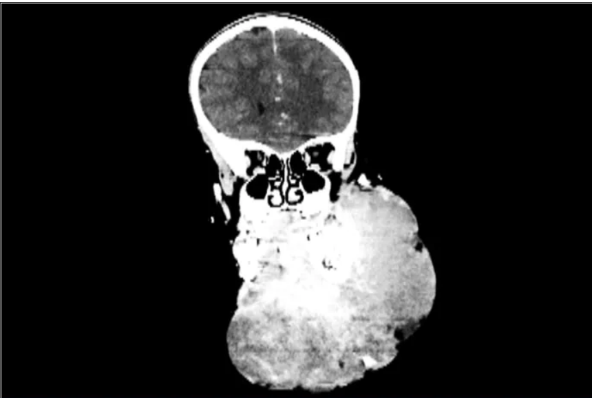
                                            An image of a scan of the patient before surgery.

The challenge with vascular malformations in children, Waner says, is that they have a fraction of the blood an adult has. Where adults have an average of 5 L of blood, a child this age has only 1 L. To lose 200 or 300 mL of blood, "that's 20 or 30% of their blood volume," Waner said. So the removal of such a mass, which requires a meticulous dissection around many blood vessels, carries a high risk of the child bleeding out.

There were some logistical hurdles, but the patient arrived in Manhattan in mid-June, at no cost to her family. The medical visa was organized by a volunteer who also work for USAID. Healing the Children Northeast paid for her travel and the Waner Kids Foundation paid for her hotel stay. Lenox Hill Hospital and Northwell Health covered all hospital costs and post-surgery care. And Drs. O and Waner did the planning, consult visits, and procedure pro bono.

The surgery was possible because of the generosity of several organizations, but the two surgeons still had a limited time to remove the mass. Under different circumstances, and with the luxury of more time, the patient would have undergone several rounds of sclerotherapy. This procedure, done by interventional radiologists, involves injecting a toxin into the blood vessels which causes them to clot. Done prior to surgery it can help limit bleeding risk.

On June 23, the morning of the surgery, the patient underwent one round of sclerotherapy. However, it didn't have the intended effect, Waner said, "because the lesion was just so massive."

The team had planned several of their moves ahead of time. But this isn't the sort of surgery you'd find in a textbook. Because it's such a unique field, Waner and O have developed many of their own techniques along the way. This patient was much like the cases they treat every day, only "several orders of magnitudes greater," Waner said. "On a scale of 1 to 10 she was a 12."


                                                    Negalem, 5, before her surgery.

The morning of the surgery, "I was very apprehensive," Waner recalled. He vividly remembers the girl's father repeatedly kissing her to say goodbye as she lay on the operating table, fully aware that this procedure was a life-threatening one. And from the beginning there were challenges, like getting her under anesthesia when the anatomy of her mouth, deformed by the tumor, didn't allow the anesthesiologists to use their typical tubing. Then, once the skin was removed, it became clear how dilated and tangled the involved blood vessels were. There were many vital structures tangled in the anomaly. "The jugular vein was right there. The carotid artery was right there," Waner said. It was extremely difficult to delineate and preserve them, he said.

Once they got into the surgery they also realized that the growth had adhered to the jaw bone. "There were vessels traversing into the bone which were hard to control," O said.

But finally, both doctors realized they'd be able to remove it. With the lesion removed they began the work of reconstruction and reanimation.

The child's jaw and cheek bone had grown beyond their normal size to support the growth. They had to shave them down to achieve facial symmetry. The tumor had also inhibited much of the child's facial nerve control. With it gone, O began the work of finding all the facial nerve branches and assembling them to reanimate the child's face.

Before medicine, O trained as an architect, which, according to Waner, has equipped her with very good spatial awareness — a valuable skill in the surgical reconstruction phase. After seeing a lecture by Waner, she immediately saw a fit for her unique interest and skill set. She did fellowship training with Waner in vascular anomalies, and then went on to specialize in facial nerve reanimation. The proof of O's expertise is Negalem's new, beautiful smile, Waner said.

The surgery drew out over 8 hours, as long as a day of surgeries for the two doctors. When O finally walked into the waiting room to inform the family of the success, the first words out of the father's mouth were, "Is my daughter alive?"

A growth like Negalem had is not compatible with a normal life. Waner's mantra is that every child has the right to look normal. But this case went beyond aesthetics. If the growth hadn't been removed, the child was only expected to live 4 to 6 more years, Waner said. Without the surgery, she could have suffocated, starved without the ability to swallow, or suffered a fatal bleed..

O and Waner are uniquely equipped to do this kind of work, but both are adamant that treating vascular anomalies is a multidisciplinary, multimodal approach. Specialties in anesthesiology, radiology, lasers, facial nerves — they are all critical to these procedures. And often patients with these kinds of lesions require medical and radiologic interventions in addition to surgery. In this particular case, from logistics to post-op, "it was a lot of teamwork," O said, "a lot of international teams coming together."

Though extremely difficult, "in the end the result was exactly what we wanted," Waner said. Negalem can live a normal life. And as for the surgical duo, both feel very fortunate to do this work. O said, "I'm honored to have found this specialty and to be able to train with and work with Milton. I'm so happy to do what I do every day."

"That's why we really took our time. We just went very slowly and deliberately," O said. The blood vessels were so dilated that their only option was to move painstakingly slow — otherwise a small nick could be devastating.

But even with the slow pace the surgery was "excruciatingly difficult," Waner said. And early on in the dissection he wasn't quite sure they'd make it out. The sclerotherapy hadn't done much to prevent bleeding. "At one point every millimeter or two that we advanced we got into some bleeding," Waner said. "Brisk bleeding."

The surgery drew out over 8 hours, as long as a day of surgeries for the two doctors. When O finally walked into the waiting room to inform the family of the success, the first words out of the father's mouth were, "Is my daughter alive?" 

A growth like Negalem had is not compatible with a normal life. Waner's mantra is that every child has the right to look normal. But this case went beyond aesthetics. If the growth hadn't been removed, the child was only expected to live 4 to 6 more years, Waner said. Without the surgery, she could have suffocated, starved without the ability to swallow, or suffered a fatal bleed.

O and Waner are uniquely equipped to do this kind of work, but both are adamant that treating vascular anomalies is a multidisciplinary, multimodal approach. Specialties in anesthesiology, radiology, lasers, facial nerves — they are all critical to these procedures. And often patients with these kinds of lesions require medical and radiologic interventions in addition to surgery. In this particular case, from logistics to post-op, "it was a lot of teamwork," O said, "a lot of international teams coming together." 

Though extremely difficult, "in the end the result was exactly what we wanted," Waner said. Negalem can live a normal life. And as for the surgical duo, both feel very fortunate to do this work. O said, "I'm honored to have found this specialty and to be able to train with and work with Milton. I'm so happy to do what I do every day." 

Donavyn Coffey is a freelance journalist who covers health and the environment from her home in the Bluegrass. Her work has appeared in Popular Science, Insider, and SELF. 

https://www.medscape.com/viewarticle/954312 

Sunday, July 11, 2021

Risk of traumatic brain injuries in infants younger than 3 months with minor blunt head trauma

Abid Z, Kuppermann N, Tancredi DJ, Dayan PS. Risk of Traumatic Brain Injuries in Infants Younger than 3 Months With Minor Blunt Head Trauma. Ann Emerg Med. 2021 Jun 17:S0196-0644(21)00298-5. doi: 10.1016/j.annemergmed.2021.04.015. Epub ahead of print. PMID: 34148662.

Abstract

Study objective: Infants with head trauma often have subtle findings suggestive of traumatic brain injury. Prediction rules for traumatic brain injury among children with minor head trauma have not been specifically evaluated in infants younger than 3 months old. We aimed to determine the risk of clinically important traumatic brain injuries, traumatic brain injuries on computed tomography (CT) images, and skull fractures in infants younger than 3 months of age who did and did not meet the age-specific Pediatric Emergency Care Applied Research Network (PECARN) low-risk criteria for children with minor blunt head trauma. 

Methods: We conducted a secondary analysis of infants <3 months old in the public use data set from PECARN's prospective observational study of children with minor blunt head trauma. Main outcomes included (1) clinically important traumatic brain injury, (2) traumatic brain injury on CT, and (3) skull fracture on CT. 

Results: Of 10,904 patients <2 years old, 1,081 (9.9%) with complete data were <3 months old; most (750/1081, 69.6%) sustained falls, and 633/1081 (58.6%) underwent CT scans. Of the 514/1081 (47.5%) infants who met the PECARN low-risk criteria, 1/514 (0.2%, 95% confidence interval [CI] 0.005% to 1.1%), 10/197 (5.1%, 2.5% to 9.1%), and 9/197 (4.6%, 2.1% to 8.5%) had clinically important traumatic brain injuries, traumatic brain injuries on CT, and skull fractures, respectively. Of 567 infants who did not meet the low-risk PECARN criteria, 24/567 (4.2%, 95% CI 2.7% to 6.2%), 94/436 (21.3%, 95% CI 17.6% to 25.5%), and 122/436 (28.0%, 95% CI 23.8% to 32.5%) had clinically important traumatic brain injuries, traumatic brain injuries, and skull fractures, respectively.

 Conclusion: The PECARN traumatic brain injury low-risk criteria accurately identified infants <3 months old at low risk of clinically important traumatic brain injuries. However, infants at low risk for clinically important traumatic brain injuries remained at risk for traumatic brain injuries on CT, suggesting the need for a cautious approach in these infants. ___________________________________________________________________

PECARN traumatic brain injury low-risk criteria for children <2 years old.

Courtesy of: https://www.mdlinx.com/journal-summary/risk-of-traumatic-brain-injuries-in-infants-younger-than-3-months-with-minor-blunt-head-trauma/57xVhgxbmFGNLSFMSZ8bGl

Tuesday, July 6, 2021

[11C]PK11195-PET brain imaging of the mitochondrial translocator protein in mitochondrial disease

Jelle van den Ameele, Young Hong, Roido Manavaki, Antonina Kouli, Heather Biggs, Zoe MacIntyre, Rita Horvath, Patrick Yu-Wai-Man, Evan Reid, Caroline H. Williams-Gray, Ed T. Bullmore, Franklin I. Aigbirhio, Tim D. Fryer, Patrick F. Chinnery. [11C]PK11195-PET Brain Imaging of the Mitochondrial Translocator Protein in Mitochondrial Disease Neurology Jun 2021, 96 (22) e2761-e2773; DOI: 10.1212/WNL.0000000000012033 

Abstract

Objective To explore the possibilities of radioligands against the mitochondrial outer membrane translocator protein (TSPO) as biomarkers for mitochondrial disease, we performed brain PET-MRI with [11C]PK11195 in 14 patients with genetically confirmed mitochondrial disease and 33 matched controls. 

Methods Case–control study of brain PET-MRI with the TSPO radioligand [11C]PK11195. 

Results Forty-six percent of symptomatic patients had volumes of abnormal radiotracer binding greater than the 95th percentile in controls. [11C]PK11195 binding was generally greater in gray matter and significantly decreased in white matter. This was most striking in patients with nuclear TYMP or mitochondrial m.3243A>G MT-TL1 mutations, in keeping with differences in mitochondrial density seen postmortem. Some regional binding patterns corresponded to clinical presentation and underlying mutation, even in the absence of structural changes on MRI. This was most obvious for the cerebellum, where patients with ataxia had decreased binding in the cerebellar cortex, but not necessarily volume loss. Overall, there was a positive correlation between aberrant [11C]PK11195 binding and clinical severity. 

Conclusion These findings endorse the use of PET imaging with TSPO radioligands as a noninvasive in vivo biomarker of mitochondrial pathology. 

Classification of Evidence This study provides Class III evidence that brain PET-MRI with TSPO radioligands identifies mitochondrial pathology.

Death by request

A University of Chicago student died Sunday, several days after being struck by a stray bullet while commuting home from an internship after reportedly signaling to his family that he wanted to be removed from life support. 

Max Soloman Lewis, 20, was commuting on the city's Green Line during rush hour on July 1 from his internship at an investment firm in downtown Chicago, the Chicago Sun-Times reported. While sitting inside the train, a bullet pierced the window and struck the back of his neck at the 51st Street/Washington Park Station, the Chicago Police Department said. 

He died Sunday morning after he was taken off life support, the newspaper reported. Authorities said Lewis wasn't the intended target and it wasn't clear where the gunfire came from. 

WGN spoke with his mother, Dr. Rebecca Rivkin, who said her son was alert, but the bullet caused catastrophic damage. He was paralyzed from the neck down and would likely need a ventilator for the rest of his life, the report said. 

He reportedly communicated with doctors and family by blinking. He spelled out on a letter board, "If I have to live like this, pull the plug please. Seriously." 

Investigators have not released a description of the shooter. 

Max Lewis, 20, a college student in Chicago, died Sunday, days after he was struck by a stray bullet while sitting inside a train during his commute home from an internship.

Max Lewis, 20, a college student in Chicago, died Sunday, days after he was struck by a stray bullet while sitting inside a train during his commute home from an internship.  (Zachary Cogan / GoFundMe) 

"Max was an intelligent, caring, compassionate member of the UChicago community and a dear friend to many," Zachary Cogan, a classmate of Lewis' and his fraternity brother, wrote on a GoFuneMe for Lewis. "He is known amongst friends, peers, and classmates for his kindness, goofiness, grit, impeccable work ethic, and most importantly, his unfailingly genuine soul. He will be sorely missed." 

"There are no words to adequately characterize how devastating this loss is," he added. "Of all people in this world, Max was the last to deserve something like this." 

Before his death, Lewis, a Denver-area native, was studying economics and computer science and was the former president of the Alpha Epsilon Pi fraternity, according to Cogan. He also devoted a fair amount of time to the Promontory Investment Research, a student group that helps other students write research reports.

https://www.foxnews.com/us/chicago-university-student-train-internship


Vagus nerve stimulation and seizure outcomes in pediatric refractory epilepsy

Jain P, Arya R. Vagus Nerve Stimulation and Seizure Outcomes in Pediatric Refractory Epilepsy: Systematic Review and Meta-Analysis. Neurology. 2021 Apr 13:10.1212/WNL.0000000000012030. doi: 10.1212/WNL.0000000000012030. Epub ahead of print. PMID: 33849993.

Abstract

Objective: We synthesized evidence for effectiveness of vagus nerve stimulation (VNS) as adjuvant therapy in pediatric drug-resistant epilepsy (DRE) by obtaining pooled estimates for seizure outcomes and analyzing their determinants. 

Methods: MEDLINE, EMBASE, and Cochrane databases were searched up to July 2019, for original research on VNS in pediatric (≤18 years-of-age) epilepsy. The primary outcome was 50% responder rate (50%-RR), the proportion of patients with ≥50% seizure reduction, at the last reported follow-up. Other outcomes included 50%-RR and proportion of seizure-free patients at additional reported time points. A random effects meta-analysis with restricted maximum likelihood estimation was performed to obtain pooled effect estimates. Meta-regression using multiple linear models was performed to obtain determinants of seizure outcomes and sources of heterogeneity. 

Results: A total of 101 studies were included. The pooled prevalence estimates for 50%-RR and seizure freedom at last follow-up (mean 2.54 years) were 56.4% (95% confidence intervals [CIs] 52.4, 60.4) and 11.6% (95% CI 9.6, 13.9) respectively. Fewer anti-seizure medications (ASMs) tried before VNS, and later age at onset of seizures were associated with better seizure outcomes following VNS implantation. An effect of sex-distribution of studies on long-term outcomes and a potential publication bias for short-term outcomes were also observed. 

Conclusion: Pooled evidence supports possible effectiveness of VNS in pediatric DRE, although complete seizure freedom is less common. Early referral (fewer trials of ASMs) may be a modifiable factor for desirable seizure outcomes with VNS from a clinical perspective.

Russo A, Hyslop A, Gentile V, Boni A, Miller I, Chiarello D, Pellino G, Zenesini C, Martinoni M, Lima M, Ragheb J, Cordelli DM, Pini A, Jayakar P, Duchowny M. Early vagus nerve stimulator implantation as a main predictor of positive outcome in pediatric patients with epileptic encephalopathy. Epileptic Disord. 2021 Jun 29. doi: 10.1684/epd.2021.1299. Epub ahead of print. PMID: 34184987.

Abstract

We describe a multicenter experience with VNS implantation in pediatric patients with epileptic encephalopathy. Our goal was to assess VNS efficacy and identify potential predictors of favorable outcome. This was a retrospective study. Inclusion criteria were: ≤18 years at the time of VNS implantation and at least one year of follow-up. All patients were non-candidates for excisional procedures. Favorable clinical outcome and effective VNS therapy were defined as seizure reduction >50%. Outcome data were reviewed at one, two, three and five years after VNS implantation. Fisher's exact test, Kaplan-Meier and multiple logistic regression analysis were employed. Twenty-seven patients met inclusion criteria. Responder rate (seizure frequency reduction ≥ 50%) at one-year follow-up was 25.9%, and 15.3% at last follow-up visit. The only variable significantly predicting favorable outcome was time to VNS implantation, with the best outcome achieved when VNS implantation was performed within five years of seizure onset (overall response rate of 83.3% at one year of follow-up and 100% at five years). In total, 63% of patients evidenced improved QOL at last follow-up visit. Only one patient exited the study due to an adverse event at two years from implantation. Early VNS implantation within five years of seizure onset was the only predictor of favorable clinical outcome in pediatric patients with epileptic encephalopathy. Improved QOL and a very low incidence of adverse events were observed.

Hereditary folate malabsorption

Huddar, Akshata, MD, DM, Chiplunkar, Shwetha, et al. Child Neurology: Hereditary Folate Malabsorption. Neurology. 2021;97(1):40-43. doi:10.1212/WNL.0000000000012083. 

Hereditary folate malabsorption (HFM, congenital folate malabsorption; OMIM#229050) is a rare, potentially treatable autosomal recessive disorder with multisystem involvement.  It is caused by homozygous or compound heterozygous mutations in SLC46A1 resulting in loss of function of proton-coupled folate transporter (PCFT),2 required for intestinal absorption and transport of folate across choroid plexus. This leads to the deficiency of folate in serum and CSF, causing hematologic, immunologic, gastrointestinal, and neurologic manifestations. Neuroimaging shows intracranial calcification. Diagnosis is confirmed by impaired absorption of an oral folate load, low CSF folate concentration (even after correction of the serum folate concentration), or the identification of pathogenic variants in SLC46A1 on molecular genetic testing.  We describe the clinical, imaging, biochemical, and genetic findings of a patient with HFM... 

Seizures became refractory to multiple antiseizure drugs at optimal dosages (phenytoin, levetiracetam, carbamazepine, topiramate, and clobazam). By 11 months of age, the patient had generalized hyperpigmentation, tremors, and oral ulcers. Developmental delay was noted at 18 months of age, with episodes of further regression following infections. The diagnosis of infantile tremor syndrome, a nutritional deficiency syndrome typically associated with vitamin B12 deficiency, characterized by pallor, developmental delay/regression, tremors, skin pigmentation, and brown scanty scalp hair, was considered at this stage. The patient received B12 supplementation. At 32 months of age, she was able to sit and stand with support, transfer objects, and say 2-3 meaningful words. There was a history of similar illness in her older sister, with seizures, fever, vomiting, loose stools, and pancytopenia, who died at 3 months of age. 

Examination revealed microcephaly (head circumference 45 cm, <3rd percentile) and low weight (10.5 kg, <3rd percentile). The patient was alert, fixed, and followed light and sound. She had normal extraocular, facial, palatal, and tongue movements. She had generalized hypotonia, normal limb movements, and brisk deep tendon reflexes. She had choreoathetosis involving distal extremities. There was no organomegaly. Fundus showed normal retina and optic discs. 

Investigations revealed anemia (hemoglobin 10.4 g/dL, reference 13-17 g/dL) with mean corpuscular volume on higher end of normal (96.6 fL, reference 83-101 fL) and normal leukocyte and platelet count. Renal and hepatic function tests were normal. Serum folate (1.52 ng/mL, reference 3.1-19.9 ng/mL) and vitamin B12 (54 pg/mL, reference 180-914 pg/mL) were low, while homocysteine was high (27.8 [mu]mol/L, reference <8 [micro]mol/L). Metabolic investigations revealed normal lactate, mildly elevated ammonia, and normal plasma amino acids, acylcarnitine profile, and urinary organic acid profile. 

Cranial CT scan done at 12 months of age was normal. MRI brain done at 13 months showed mild cerebral atrophy and hypointensity involving the basal ganglia and left parieto-occipital region on T1-weighted images  with corresponding hyperintensity on T2-weighted sequences. Repeat neuroimaging at 32 months of age showed progressive changes, including calcification in bilateral basal ganglia and thalamus on CT scan. MRI brain done at 32 months showed diffuse cerebral and cerebellar atrophy, T2/fluid-attenuated inversion recovery hyperintensities of periventricular and deep white matter, and mixed intensity lesions in bilateral basal ganglia. EEG showed focal epileptiform activity across the left temporal region. 

Constellation of megaloblastic anemia, oral ulcers, recurrent infections, and neurologic features with positive family history and biochemical and imaging findings led to the suspicion of hereditary folate deficiency. CSF folate estimation was not done. Clinical exome sequencing showed a pathogenic homozygous single base pair duplication in exon 2 of the SLC46A1 (chr17:26732094_26732095dupC) c.620dupG variation resulting in a frameshift and premature truncation of the protein 25 amino acids downstream to codon 208 (p. Tyr208LeufsTer25; ENST00000440501), confirming the diagnosis of HFM. Both parents were heterozygous carriers. 

The patient received oral folic acid from 4 months of age prior to presentation to our facility. Hematologic measures and hyperpigmentation improved, but neurologic features worsened, and she continued to have recurrent infections. After the genetic diagnosis, she was treated with intramuscular injection of folinic acid (l20 mg/d), leading to the cessation of seizures. After 1 week, parenteral folinic acid was replaced by oral folinic acid (150 mg/d, 15 mg/kg/d) due to financial constraints and continued along with anticonvulsants. At 2 months follow-up, she was seizure-free, and development had stabilized. However, she was noncompliant with treatment and presented multiple times with recurrent seizures, anemia, and infections, which responded to parenteral folinic acid. On follow-up at 7 years, the patient was able to stand with support, speak 5-10 words, and feed herself. Although there was a reduction in seizure frequency, she continued to have daily seizures requiring multiple anticonvulsants.

The patient received oral folic acid from 4 months of age prior to presentation to our facility. Hematologic measures and hyperpigmentation improved, but neurologic features 

MEHMO syndrome

Moortgat S, Manfroid I, Pendeville H, Freeman S, Bourdouxhe J, Benoit V, Merhi A, Philippe C, Faivre L, Maystadt I. Broadening the phenotypic spectrum and physiological insights related to EIF2S3 variants. Hum Mutat. 2021 Jul;42(7):827-834. doi: 10.1002/humu.24215. Epub 2021 May 19. PMID: 33942450.

Abstract

Mental deficiency, epilepsy, hypogonadism, microcephaly, and obesity syndrome is a severe X-linked syndrome caused by pathogenic variants in EIF2S3. The gene encodes the γ subunit of the eukaryotic translation initiation factor-2, eIF2, essential for protein translation. A recurrent frameshift variant is described in severely affected patients while missense variants usually cause a moderate phenotype. We identified a novel missense variant (c.433A>G, p.(Met145Val)) in EIF2S3 in a mildly affected patient. Studies on zebrafish confirm the pathogenicity of this novel variant and three previously published missense variants. CRISPR/Cas9 knockout of eif2s3 in zebrafish embryos recapitulate the human microcephaly and show increased neuronal cell death. Abnormal high glucose levels were identified in mutant embryos, caused by beta cell and pancreatic progenitor deficiency, not related to apoptosis. Additional studies in patient-derived fibroblasts did not reveal apoptosis. Our results provide new insights into disease physiopathology, suggesting tissue-dependent mechanisms.

Skopkova M, Hennig F, Shin BS, Turner CE, Stanikova D, Brennerova K, Stanik J, Fischer U, Henden L, Müller U, Steinberger D, Leshinsky-Silver E, Bottani A, Kurdiova T, Ukropec J, Nyitrayova O, Kolnikova M, Klimes I, Borck G, Bahlo M, Haas SA, Kim JR, Lotspeich-Cole LE, Gasperikova D, Dever TE, Kalscheuer VM. EIF2S3 Mutations Associated with Severe X-Linked Intellectual Disability Syndrome MEHMO. Hum Mutat. 2017 Apr;38(4):409-425. doi: 10.1002/humu.23170. Epub 2017 Jan 23. PMID: 28055140; PMCID: PMC6267786.

Abstract

Impairment of translation initiation and its regulation within the integrated stress response (ISR) and related unfolded-protein response has been identified as a cause of several multisystemic syndromes. Here, we link MEHMO syndrome, whose genetic etiology was unknown, to this group of disorders. MEHMO is a rare X-linked syndrome characterized by profound intellectual disability, epilepsy, hypogonadism and hypogenitalism, microcephaly, and obesity. We have identified a C-terminal frameshift mutation (Ile465Serfs) in the EIF2S3 gene in three families with MEHMO syndrome and a novel maternally inherited missense EIF2S3 variant (c.324T>A; p.Ser108Arg) in another male patient with less severe clinical symptoms. The EIF2S3 gene encodes the γ subunit of eukaryotic translation initiation factor 2 (eIF2), crucial for initiation of protein synthesis and regulation of the ISR. Studies in patient fibroblasts confirm increased ISR activation due to the Ile465Serfs mutation and functional assays in yeast demonstrate that the Ile465Serfs mutation impairs eIF2γ function to a greater extent than tested missense mutations, consistent with the more severe clinical phenotype of the Ile465Serfs male mutation carriers. Thus, we propose that more severe EIF2S3 mutations cause the full MEHMO phenotype, while less deleterious mutations cause a milder form of the syndrome with only a subset of the symptoms.

Steinmüller R, Steinberger D, Müller U. MEHMO (mental retardation, epileptic seizures, hypogonadism and -genitalism, microcephaly, obesity), a novel syndrome: assignment of disease locus to xp21.1-p22.13. Eur J Hum Genet. 1998 May-Jun;6(3):201-6. doi: 10.1038/sj.ejhg.5200180. PMID: 9781023.

Abstract

A previously unrecognised X-chromosomal mental retardation syndrome is described. Clinical hallmarks are mental retardation, epileptic seizures, hypogonadism, and -genitalism, microcephaly and obesity. Life expectancy of patients is less than two years. Based on the major clinical symptoms this condition is referred to by the acronym MEHMO. Haplotype and two-point linkage analyses in a large three-generation family assign the disease locus to Xp21.1-p22.13, to a region that is flanked by CYBB and DXS365.

Leshinsky-Silver E, Zinger A, Bibi CN, Barash V, Sadeh M, Lev D, Sagie TL. MEHMO (Mental retardation, Epileptic seizures, Hypogenitalism, Microcephaly, Obesity): a new X-linked mitochondrial disorder. Eur J Hum Genet. 2002 Apr;10(4):226-30. doi: 10.1038/sj.ejhg.5200791. PMID: 12032729.

Abstract

MEHMO (Mental retardation, Epileptic seizures, Hypogenitalism, Microcephaly and Obesity) is an X-linked disorder characterised by mental retardation, epileptic seizures, hypogenitalism, microcephaly and obesity. It was recently assigned to the locus Xp21.1-p22.13. We describe a child with MEHMO and lactic acidosis whose muscle biopsy revealed markedly reduced activities of complexes 1,3 and 4 of the mitochondrial electron transport chain. Histological staining showed mitochondrial proliferation and lipid storage. Electron microscopy revealed abnormal and enlarged mitochondria with concentric cristae and electron dense bodies. This is the first identification of MEHMO as a mitochondrial disorder and one of the very few X-linked mitochondrial syndromes.