Thursday, February 28, 2019

Behind the seizure


Eligibility for a program that provides free genetic testing for epilepsy in young children has been expanded. The Behind the Seizure program, which had been available for patients age 2 to 4 years with a history of 1 or more unprovoked seizures, is now offered for children from birth to age 5 years.

Over the past 2 years, hundreds of children have received testing through this program, and research has shown that participants were diagnosed 1 to 2 years sooner than historic averages for the diagnoses identified.

The 4 corporate partners in the program (Invitae Corporation, Stoke Therapeutics, Xenon Pharmaceuticals, and BioMarin Pharmaceutical sponsor the cost of testing a gene panel (Invitae Epilepsy Panel; Invitae Corporation, San Francisco, CA) of over 180 genes associated with both syndromic and non-syndromic causes of epilepsy, including neurodegenerative conditions.

On average, test results are available in 14 days. "Through this program we can secure comprehensive genetic testing for a child who has had an unprovoked seizure at no cost to the patient,” said Raman Sankar, MD, professor and chief of pediatric neurology at UCLA Mattel Children's Hospital in Los Angeles. “That testing helps us get to a diagnosis faster so we can focus on providing the most accurate and timely treatment for our patients. When dealing with such young patients, this type of program can be very important, shortening the time to diagnosis, particularly for neurodegenerative conditions such as CLN2 disease."


No-cost epilepsy gene panel testing program

Invitae, BioMarin, Stoke Therapeutics, and Xenon Pharmaceuticals have partnered to offer Invitae's comprehensive epilepsy panel to any child up to 60 months old who has had an unprovoked seizure.

An epilepsy gene panel can bring you closer to understanding what’s causing your patient’s epilepsy, helping your patients and their caregivers benefit from deeper knowledge and timely care.

More than 50% of epilepsies have some genetic basis. When a patient presents with seizures, genetic testing can help identify 100+ underlying genetic causes. Early genetic testing may be the most direct, cost-effective, and accurate diagnostic tool.1

Many genes are actionable. Gene panels increasingly help tailor your approach—more than 20 genes have been linked to specific treatment strategies. Identifying a seizure’s underlying etiology can enable more precise treatment.

Delay can be devastating for patients with genetic epilepsy. Some genetic epilepsies are neurodegenerative, and may initially present with subtle, non-specific symptoms like language development delay and/ or motor disturbance. Genetic testing can help identify the cause of epilepsy before a patient experiences the hallmark signs of regression.

Genetic epilepsies may be hiding behind non-specific symptoms. Language delay and motor disturbance may be the best predictors of finding genetic epilepsies.

https://www.invitae.com/en/behindtheseizure/

I know that I matter


See: https://childnervoussystem.blogspot.com/2015/05/triumph-over-adversity.html?showComment=1431617658913#c912397699770123724
 https://childnervoussystem.blogspot.com/2016/01/triumph-over-adversity-2.html?showComment=1453336299938#c5307845136266627846
https://childnervoussystem.blogspot.com/2016/01/triumph-over-adversity-2.html?showComment=1459448449126#c789168220191724973

Wednesday, February 27, 2019

Intravenous metoclopramide in the treatment of acute migraines


Doğan NÖ, Pekdemir M, Yılmaz S, Yaka E, Karadaş A, Durmuş U, Avcu N, Koçkan E. Intravenous metoclopramide in the treatment of acute migraines: A randomized, placebo-controlled trial.  Acta Neurol Scand. 2019 Jan 10. doi: 10.1111/ane.13063. [Epub ahead of print]

Abstract

Objectives
The present study aimed to evaluate the efficacy and safety of intravenous metoclopramide for acute migraine treatment.

Materials and methods
A double‐blind, randomized, parallel‐group, placebo‐controlled trial was carried out in an academic emergency department. After the patients were assessed for eligibility via the International Headache Society criteria for migraines, they were randomized into 10 mg intravenous metoclopramide and normal saline groups. The headache intensity was evaluated using an 11‐point numeric rating scale (NRS) score. The primary outcome measure was determined as the median between‐group change in the score at the 30th minute. The secondary outcome measures were rescue medication needs, adverse events, and emergency department (ED) revisits after discharge.

Results
A total of 148 patients were randomized into two equal groups with similar baseline characteristics, including the baseline NRS scores (8 points). The median reduction in the NRS scores at the 30th minute was 4 [interquartile range (IQR): 2‐6)] in the metoclopramide group and 3 (IQR: 1‐4) in the normal saline group [median difference: −1.0, 95% confidence interval (CI): −2.1 to 0.1]. No serious adverse events were observed, and the rescue medication needs were similar in both groups.

Conclusion
No difference was found between intravenous metoclopramide and placebo regarding efficacy and safety in patients with acute migraines.

Tuesday, February 26, 2019

Pediatric myotonic dystrophy


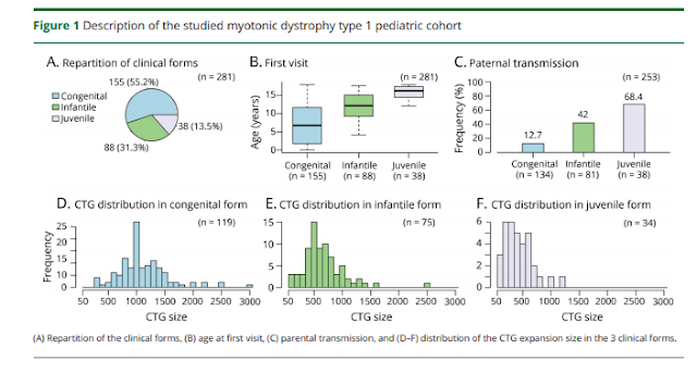
Lagrue E, Dogan C, De Antonio M, Audic F, Bach N, Barnerias C, Bellance R, Cances C, Chabrol B, Cuisset JM, Desguerre I, Durigneux J, Espil C, Fradin M, Héron D, Isapof A, Jacquin-Piques A, Journel H, Laroche-Raynaud C, Laugel V, Magot A, Manel V, Mayer M, Péréon Y, Perrier-Boeswillald J, Peudenier S, Quijano-Roy S, Ragot-Mandry S, Richelme C, Rivier F, Sabouraud P, Sarret C, Testard H, Vanhulle C, Walther-Louvier U, Gherardi R, Hamroun D, Bassez G. A large multicenter study of pediatric myotonic dystrophy type 1 for evidence-based management. Neurology. 2019 Feb 19;92(8):e852-e865.

Abstract

OBJECTIVE:
To genotypically and phenotypically characterize a large pediatric myotonic dystrophy type 1 (DM1) cohort to provide a solid frame of data for future evidence-based health management.

METHODS:
Among the 2,697 patients with genetically confirmed DM1 included in the French DM-Scope registry, children were enrolled between January 2010 and February 2016 from 24 centers. Comprehensive cross-sectional analysis of most relevant qualitative and quantitative variables was performed.

RESULTS:
We studied 314 children (52% females, with 55% congenital, 31% infantile, 14% juvenile form). The age at inclusion was inversely correlated with the CTG repeat length. The paternal transmission rate was higher than expected, especially in the congenital form (13%). A continuum of highly prevalent neurodevelopmental alterations was observed, including cognitive slowing (83%), attention deficit (64%), written language (64%), and spoken language (63%) disorders. Five percent exhibited autism spectrum disorders. Overall, musculoskeletal impairment was mild. Despite low prevalence, cardiorespiratory impairment could be life-threatening, and frequently occurred early in the first decade (25.9%). Gastrointestinal symptoms (27%) and cataracts (7%) were more frequent than expected, while endocrine or metabolic disorders were scarce.

CONCLUSIONS:
The pedDM-Scope study details the main genotype and phenotype characteristics of the 3 DM1 pediatric subgroups. It highlights striking profiles that could be useful in health care management (including transition into adulthood) and health policy planning.


Monday, February 25, 2019

Moving toward “virtual biopsy” of gliomas using artificial intelligence


What if you could know, from a pre-operative MRI, whether a glioma was likely to have a genetic mutation that significantly alters prognosis? 

Preliminary success in using machine learning to analyze MRI data suggests the promise of artificial intelligence (AI) to create a “virtual biopsy” that would increase pre-operative precision in diagnosis and prognosis of brain tumors, including glioblastoma multiforme (GBM).

Earlier this year, a research team that included Omar Arnaout, MD, a neurosurgeon in the Brigham and Women’s Hospital Department of Neurosurgery, demonstrated the use of AI to identify isocitrate dehydrogenase (IDH) mutation based on MRI data. IDH mutation, which confers significantly longer survival, was chosen for testing because IDH status may influence treatment plans, perioperative patient counseling and adjuvant management.

“Historically, we’ve needed tissue. Now, we can predict, very reliably just from an MRI, whether the patient has this mutation. Theoretically, that can guide your conversation with the patient, guide your surgical aggressiveness, and guide the kind of treatment,” said Dr. Arnaout, co-director of the Computational Neuroscience Outcomes Center (CNOC) @Harvard, which focuses on neurosurgical applications of artificial intelligence. The Center is based in Brigham and Women’s Department of Neurosurgery.

Arnaout and a team from Brigham and Women’s Hospital, along with colleagues from other hospitals, tested whether the IDH status of gliomas could be predicted accurately from MR imaging by applying a residual convolutional neural network to preoperative radiographic data. To do this, the researchers combined preoperative imaging from 496 patients at three hospitals (including the Brigham) and used two patient cohorts to train a machine to differentiate IDH-mutated tumors (associated with longer overall survival) from tumors without the mutation (which have a poor prognosis). Independent performance testing on a third cohort predicted IDH status with 79 to 87 percent accuracy, the authors reported in Clinical Cancer Research.

Building on that foundation, Arnaout and colleagues at Brigham and Women’s Hospital, working closely with their collaborators, are conducting prospective validation of pre-operative mutation detection in gliomas. This moves them closer to their goal of creating easily-usable “virtual biopsy” software – with broad implications.

“IDH is only one of many clinically relevant mutations in GBM,” Arnaout said. “We’re taking structural MRI data and using it to predict not only biomarker status in a brain tumor but also clinical outcome and use that as a decision aid.”

The long history of neurosurgery at Brigham and Women’s Hospital and its large volume of patient data uniquely positions Arnaout and his CNOC colleagues. Ongoing work combines multiple rich data sets to contribute to machine learning:

-Images, including perioperative CT and MRI and from post-surgical monitoring
-“Unstructured data” from electronic medical records, analyzed through natural language processing
-Pathology, dating back several decades at Brigham and Women’s, now being digitized and used as input for machine learning algorithms
-Physiological data from patients in Brigham and Women’s Neurosurgical Intensive Care Unit that is being collected and archived for research

“AI can be a good tool for balancing treatment effectiveness versus patient harm for each individual case. This can help us focus the difficult conversations around risk and benefit, to optimize quality of life,” said Arnaout.

Another CNOC initiative, led by CNOC co-director Timothy R. Smith, MD, PhD, MPH, involves collecting data outside the context of the healthcare environment to help predict readmissions and to identify early any tumor recurrence or decline in cognitive function. This digital phenotyping includes tracking smart-phone data of patients to assess, from their daily usage, whether they are experiencing subtle cognitive decline.

Multiple review articles published in 2018 by Drs. Arnaout and Smith, with CNOC co-director William Brian Gormley, MD, MPH, MBA, MBA and other colleagues offer an introduction and overview and the potential for outcome prediction in use of AI in neurosurgery.

https://brighamhealthvitallines.org/2018/12/18/moving-toward-virtual-biopsy-of-gliomas-using-artificial-intelligence/?utm_source=linkedin&utm_medium=social&utm_campaign=2019usnp&utm_content=neuro_c2

Sunday, February 24, 2019

Medication error- mistake or homicide?


A Tennessee nurse charged with reckless homicide after a medication error killed a patient pleaded not guilty on Wednesday in a Nashville courtroom packed with other nurses who came in scrubs to show their support.

The error happened at Vanderbilt University Medical Center in December 2017 when RaDonda Vaught injected 75-year-old Charlene Murphey with the paralytic vecuronium instead of the sedative Versed.

The 35-year-old Vaught could not find Versed in an automatic dispensing cabinet, so she used an override mechanism to type in "VE" then picked the first drug that came up, according to court documents and a report from the Centers for Medicare and Medicaid Services.

Speaking to reporters after Wednesday's hearing, Vaught's attorney, Peter Strianse, called the criminal charge against the nurse "completely unfathomable." He noted that the state board of nursing has taken no action against Vaught's nursing license, which is still active.

"This sets a terrible precedent, and these nurses are here today because it is patently unfair to charge a nurse with a criminal offense for something that was nothing more than a mistake," Strianse said.

Vaught did not want to discuss the case, but she and several of the other nurses teared up as she spoke of the "overwhelming" support she has received.

"I'm very thankful that I picked a profession with such generous, loving people," Vaught said. Online supporters have donated more than $72,000 for her legal bills.

Murphey's husband, Sam Murphey, reached by phone said he is too upset to talk about his wife. Her son, Gary Murphey, told The Tennessean newspaper earlier this month that his mother would have forgiven Vaught and felt sorrow for her.

"I know my mom well, and she would be very upset knowing that this lady may spend some of her life in prison," Gary Murphey told The Tennessean. "She probably had a family, and it's destroyed their life too."

He also said the family has no plans to sue the hospital.

Janie Harvey Garner runs the online nurse advocacy organization Show Me Your Stethoscope. She has been organizing support for Vaught and flew in from St. Louis for the hearing.

Garner said medication errors happen all the time but usually the public is unaware of them. And she said nurses don't take the errors lightly.

"RaDonda has to wake up every day and think about what happened," Garner said. "I, early in my career, made a minor error. No one was harmed. But every time I think about it, I sweat."

Garner said she believes Murphey's death was terrible and tragic. But she worries Vaught's prosecution will ultimately hurt patient safety.

"This will cause people to die, because people won't come forward with their mistakes," Garner said…

Nurse Marguerite McBride was at the Wednesday hearing to support Vaught and said she had worked with her at a different hospital for about a year.

"She's an amazing, compassionate, caring nurse," McBride said. "Families love her. Other nurses love her."

https://www.foxnews.com/us/nurse-charged-in-fatal-drug-swap-error-pleads-not-guilty

Courtesy of a colleague

See: https://childnervoussystem.blogspot.com/2016/05/medical-error.html
https://childnervoussystem.blogspot.com/2018/02/to-err-is-homicide-in-britain.html 

Saturday, February 23, 2019

ADEM-clinical and MRI decision making in the emergency department


Orly Bisker Kassif, Rotem Orbach, Ayelet Rimon, Dennis Scolnik, Miguel Glatstein.  Acute disseminated encephalomyelitis in children - clinical and MRI decision making in the emergency department.  The American Journal of Emergency Medicine.  In press.

Abstract

Background
Acute disseminated encephalomyelitis (ADEM) is an uncommon, treatable, primarily pediatric, immune-mediated disease. Diagnosis of ADEM requires two essential elements: typical clinical presentation and magnetic resonance imaging (MRI) findings. The aim of this study was to evaluate how clinical findings in the initial emergency department (ED) presentation influenced the timing of MRI.

Methods
A retrospective chart review was conducted of children diagnosed with ADEM, over a 12-year period, in a tertiary care pediatric center. Clinical presentation at ED admission was recorded and patients who underwent an MRI as part of their ED evaluation (early MRI) with those who had MRI performed during ward hospitalization (late MRI) were compared.

Results
30 patients were diagnosed with ADEM during the study period. Encephalopathy and polyfocal neurological signs were described in 80% and 50% of patients ED charts, respectively. Seven patients underwent early MRI and polyfocal neurological signs were more common in this group (p = 0.006). Fever was more common in the late MRI group (p = 0.02). Following diagnosis, all patients were treated with immune-modulation therapy, improved clinically, and were discharged.

Conclusion
20% of ADEM patients were not encephalopathic at ED presentation. Polyfocal neurological signs and absence of fever at ED presentation were related to earlier MRI utilization and thus earlier diagnosis and treatment. Familiarity with the ADEM constellation of signs, and a high index of suspicion, may help the ED clinician in early diagnosis and treatment of this rare disease.

Thursday, February 21, 2019

China’s CRISPR twins might have had their brains inadvertently enhanced

The brains of two genetically edited girls born in China last year may have been changed in ways that enhance cognition and memory, scientists say.

The twins, called Lulu and Nana, reportedly had their genes modified before birth by a Chinese scientific team using the new editing tool CRISPR. The goal was to make the girls immune to infection by HIV, the virus that causes AIDS.

Now, new research shows that the same alteration introduced into the girls’ DNA, to a gene called CCR5, not only makes mice smarter but also improves human brain recovery after stroke, and could be linked to greater success in school.

“The answer is likely yes, it did affect their brains,” says Alcino J. Silva, a neurobiologist at the University of California, Los Angeles, whose lab uncovered a major new role for the CCR5 gene in memory and the brain’s ability to form new connections.

“The simplest interpretation is that those mutations will probably have an impact on cognitive function in the twins,” says Silva. He says the exact effect on the girls’ cognition is impossible to predict, and “that is why it should not be done.”

he Chinese team, led by He Jiankui of the Southern University of Science and Technology in Shenzhen, claimed it used CRISPR to delete CCR5 from human embryos, some of which were later used to create pregnancies. HIV requires the CCR5 gene to enter human blood cells.

The experiment has been widely condemned as irresponsible, and He is under investigation in China. News of the first gene-edited babies also inflamed speculation about whether CRISPR technology could one day be used to create super-intelligent humans, perhaps as part of a biotechnology race between the US and China.

There is no evidence that He actually set out to modify the twins’ intelligence. MIT Technology Review contacted scientists studying the effects of CCR5 on cognition, and they say the Chinese scientist never reached out to them, as he did to others from whom he hoped to get scientific advice or support.

“As far as I know, we never heard from him,” says Miou Zhou, a professor at the Western University of Health Sciences in California.

Although He never consulted the brain researchers, the Chinese scientist was certainly aware of link between CCR5 and cognition. It was first shown in 2016 by Zhou and Silva, who found that removing the gene from mice significantly improved their memory. The team had looked at more than 140 different genetic alterations to find which made mice smarter.

Silva says because of his research, he sometimes interacts with figures in Silicon Valley and elsewhere who have, in his opinion, an unhealthy interest in designer babies with better brains. That’s why, when the birth of the twins became public on November 25, Silva says he immediately wondered if it had been an attempt at this kind of alteration. “I suddenly realized—Oh, holy shit, they are really serious about this bullshit,” says Silva. “My reaction was visceral repulsion and sadness.”

Whatever He’s true aims, evidence continues to build that CCR5 plays a major role in the brain. Today, for example, Silva and a large team from the US and Israel say they have new proof that CCR5 acts as a suppressor of memories and synaptic connections.

According to their new report, appearing in the journal Cell, people who naturally lack CCR5 recover more quickly from strokes. What’s more, people missing at least one copy of the gene seem to go further in school, suggesting a possible role in everyday intelligence.

“We are the first to report a function of CCR5 in the human brain, and the first to report a higher level of education,” says UCLA biologist S. Thomas Carmichael, who led the new study. He calls the link to educational success “tantalizing” but says it needs further study.

The discoveries about CCR5 are already being followed up in drug trials on both stroke patients and people with HIV, who sometimes suffer memory problems. In those studies, one of which is under way at UCLA, people are being given an anti-HIV drug, Maraviroc, which chemically blocks CCR5, to see if it improves their cognition.

Silva says there is a big difference between trying to correct deficits in such patients and trying to create enhancement. “Cognitive problems are one of the biggest unmet needs in medicine. We need drugs, but it’s another thing to take normal people and muck with the DNA or chemistry to improve them,” he says. “We simply don’t know enough to do it. Nature has struck a very fine balance.”

Just because we shouldn’t alter normal intelligence doesn’t mean we can’t. Silva says the genetic manipulations used to make “smart mice” show not only that it is possible, but that changing CCR5 has particularly big effects.

“Could it be conceivable that at one point in the future we could increase the average IQ of the population? I would not be a scientist if I said no. The work in mice demonstrates the answer may be yes,” he says. “But mice are not people. We simply don’t know what the consequences will be in mucking around. We are not ready for it yet.”
_______________________________________________________________________

CCR5 Is a Therapeutic Target for Recovery after Stroke and Traumatic Brain Injury
Mary T. Joy, Einor Ben Assayag, Dalia Shabashov-Stone, Alcino J. Silva, Esther Shohami, S. Thomas Carmichael.  CCR5 Is a Therapeutic Target for Recovery after Stroke and Traumatic Brain Injury.  Cell. 2019 Feb 21; 176(5):1143-1157.E13.
Highlights
CCR5 is differentially upregulated in neurons after stroke
Knockdown of CCR5 induces motor recovery after stroke and improves cognition after TBI
Treatment with an FDA-approved drug, maraviroc induces recovery after stroke and TBI
Human carriers for CCR5delta32 have better outcomes after stroke

Summary
We tested a newly described molecular memory system, CCR5 signaling, for its role in recovery after stroke and traumatic brain injury (TBI). CCR5 is uniquely expressed in cortical neurons after stroke. Post-stroke neuronal knockdown of CCR5 in pre-motor cortex leads to early recovery of motor control. Recovery is associated with preservation of dendritic spines, new patterns of cortical projections to contralateral pre-motor cortex, and upregulation of CREB and DLK signaling. Administration of a clinically utilized FDA-approved CCR5 antagonist, devised for HIV treatment, produces similar effects on motor recovery post stroke and cognitive decline post TBI. Finally, in a large clinical cohort of stroke patients, carriers for a naturally occurring loss-of-function mutation in CCR5 (CCR5-Δ32) exhibited greater recovery of neurological impairments and cognitive function. In summary, CCR5 is a translational target for neural repair in stroke and TBI and the first reported gene associated with enhanced recovery in human stroke.

See:  https://childnervoussystem.blogspot.com/2018/11/gene-edited-babies.html

Treatment of infantile spasms 2

Hussain SA. Treatment of infantile spasms. Epilepsia Open. 2018 Oct 23;3(Suppl Suppl 2):143-154.

Abstract
The treatment of infantile spasms is challenging, especially in the context of the following: (1) a severe phenotype with high morbidity and mortality; (2) the urgency of diagnosis and successful early response to therapy; and (3) the paucity of effective, safe, and well-tolerated therapies. Even after initially successful treatment, relapse risk is substantial and the most effective therapies pose considerable risk with long-term administration. In evaluating any treatment for infantile spasms, the key short-term outcome measure is freedom from both epileptic spasms and hypsarrhythmia. In contrast, the most important long-term outcomes are enduring seizure-freedom and measures of intellectual performance in later childhood and adulthood. First-line treatment options-namely hormonal therapy and vigabatrin-display moderate to high efficacy but also exhibit substantial side-effect burdens. Data on efficacy and safety of each class of therapy, as well as the combination of these therapies, are reviewed in detail. Specific hormonal therapies (adrenocorticotropic hormone and various corticosteroids) are contrasted. Those etiologies that prompt specific therapies are reviewed briefly, as are an array of second-line therapies supported by less-compelling data. The ketogenic diet is discussed in greater detail, with a focus on the limitations of numerous available studies that generally suggest that it is efficacious. Special discussion is allocated to cannabidiol-the investigational therapy that has received the most attention, and which is already in use in the form of various artisanal cannabis extracts. Finally, a treatment algorithm reflecting the concepts and controversies discussed in this review is presented.

Treatment of acute migraine by a partial rebreathing device


Fuglsang CH, Johansen T, Kaila K, Kasch H, Bach FW. Treatment of acute migraine by a partial rebreathing device: A randomized controlled pilot study. Cephalalgia. 2018 Sep;38(10):1632-1643.

Abstract
Background Impaired brain oxygen delivery can trigger and exacerbate migraine attacks. Normoxic hypercapnia increases brain oxygen delivery markedly by vasodilation of the cerebral vasculature, and hypercapnia has been shown to abort migraine attacks. Stable normoxic hypercapnia can be induced by a compact partial rebreathing device. This pilot study aimed to provide initial data on the device's efficacy and safety. Methods Using a double-blinded, randomized, cross-over study design, adult migraine-with-aura patients self-administered the partial rebreathing device or a sham device for 20 minutes at the onset of aura symptoms. Results Eleven participants (mean age 35.5, three men) self-treated 41 migraine attacks (20 with the partial rebreathing device, 21 with sham). The partial rebreathing device increased mean End Tidal CO2 by 24%, while retaining mean oxygen saturation above 97%. The primary end point (headache intensity difference between first aura symptoms and two hours after treatment (0-3 scale) - active/sham difference) did not reach statistical significance (-0.55 (95% CI: -1.13-0.04), p = 0.096), whereas the difference in percentage of attacks with pain relief at two hours was significant ( p = 0.043), as was user satisfaction ( p = 0.022). A marked efficacy increase was seen from first to second time use of the partial rebreathing device. No adverse events occurred, and side effects were absent or mild. Conclusion Normoxic hypercapnia shows promise as an adjunctive/alternative migraine treatment, meriting further investigation in a larger population. Clinical study registered at ClinicalTrials.gov with identifier NCT03472417.

Courtesy of:  https://www.medscape.com/viewarticle/908009?nlid=127889_3001&src=WNL_mdplsfeat_190212_mscpedit_neur&uac=60196BR&spon=26&impID=1882939&faf=1


Wednesday, February 20, 2019

PANS and PANDAS

Inspired by a colleague

PANS AND PANDAS: AN INTERVIEW WITH DR. MIROSLAV KOVACEVIC
9/4/2018 

Dr. Kovacevic is a board certified pediatrician and one of the first pediatricians in the country to begin treating children with PANDAS.
How did you first learn of PANS/PANDAS and what motivated you to begin treating children with PANS and PANDAS?

It was an accident. In 1999 a child of a friend, who was a pediatrician, had a condition nobody could explain so that is how it started. I found a small five liner in the literature referring to PANDAS and tried to contact the NIH. I did get in touch with them and was told they were doing some research on it. After that, I had kids were so incapacitated and had been through five or six institutions. I tried treatment for PANDAS, it worked, and from them on I continued to treat children who had PANDAS.

Do you ever treat children who did not have an abrupt or acute onset, and if so, do they respond similarly to children who did have an abrupt onset?

There has been an evolution of understanding of PANS and PANDAS. The first ten to twelve years, the sudden onset was a must. When a child starts having symptoms at two to three years of age, it is more difficult to recognize the onset. Certainly there are children who fulfill all the criteria aside from abrupt onset for whom treatment is successful.

Have you had any patients who had been diagnosed with autism who ended
up actually having PANS?

Yes, but let’s be specific on that. I have had a number of children who were initially diagnosed with PDD-NOS that later on turned out to be PANDAS.

And did their symptoms of autism remit with treatment for PANDAS?

Yes, their symptoms of autism went away with treatment.

What about children who had been diagnosed with bipolar disorder, oppositional defiant disorder, etc?

Any time you have children with a long history of neuropsychiatric symptoms, you will have a whole slew of diagnoses--anxiety, oppositional defiant disorder, bipolar disorder, etc. That really has been the story. You have all these diagnoses established at different times. The symptoms evolution of PANDAS over time differs from child to child. A child can have one episode where OCD is the main feature. The next episode could be tics or irrational fears. Children's symptoms do change from episode to episode and advance with age.

What are the ages of the youngest and the oldest PANS patients you’ve ever diagnosed?

The youngest patient with confirmed PANDAS was 3.5 years old and presented with acute anorexia. The oldest was 48. Up until 2010, PANDAS was considered to be exclusively pediatric. I started to question this and found that these children do not outgrow PANDAS. It doesn’t go away so they retain the symptoms. It appears that unless children with PANDAS are treated, it is a lifelong condition.

In your opinion, why hasn’t PANS/PANDAS moved beyond controversy?

I think recently, as recently as the past two to three years, it has moved beyond controversy. I think one of the reasons it has been difficult is we still have the division in medicine into mental and physical symptoms. There is mental illness which in my opinion doesn’t exist. There is physical illness with mental illness symptoms. Children with PANDAS often have no physical symptoms of illness so they’re immediately pronounced behavioral. There is the  implication that the child could do something about their behavior if they tried hard enough. That is unfair.

The second problem is there is a division between mental and physical illness that is deeply rooted into society. There are two groups of patients and they are treated differently. In mental illness, there is usually not normal testing, lab testing, etc. that is required in physical illness. Again, switching camps is very hard.

What percentage of your patient population requires IVIG?

Again it is a matter of age. In my opinion, and I have followed patients as long as 19 years, it appears all patients with a diagnosis of PANDAS eventually do need IVIG as the ultimate
resolution. Yes, you can put these patients into remission with antibiotics and steroids, however, based on my experience, the occurrence is almost inevitable later in life. Let me give you an example. A number of years ago, I had a child with acute onset at eight years old, returning from Europe. We treated with antibiotics and he did well. It happened that I followed the child as pediatrician at that time. I was witness to his perfectly normal health for five to six years. At age 14, he woke up with full blown picture of PANDAS. I believe all children with PANDAS will need IVIG sooner or later.

How often is more than one course of ivig necessary?

Again historically, it all depends on what is done prior to IVIG. Until 2010 we basically would treat with IVIG if the child's clinical picture required it. I saw a 18-20% recurrence. In 2010 we started look at recurrence more closely and tonsils and adenoids were suspected as a trigger.  We started to employ tonsillectomy and adenoidectomy before treating with IVIG. Preliminary evidence is 5% or less are now needing repeat IVIG. Somewhere between
one in ten and one in twenty children.

Do you see a difference in outcomes when children are treated promptly
versus when they have been sick for years?

Actually that is not necessarily the case. It appears the effectiveness is solely dependent upon the age, not the duration, intensity, or quality of symptoms. From my population, in boys between the ages of six and 13, and girls between the ages of six and 12 who are treated with IVIG, the response rate appears to be 75-77%. After twelve in boys and thirteen in girls,the effectiveness starts slowly waning down. The effectiveness dissipates entirely by late twenties or early thirties. The importance of early diagnosis is not necessarily related
to better outcomes.  Clearly improving quality of life for children and parents because they know what they are dealing with is an importance that should be attached to early diagnosis.

Is there any new research you're excited about that you think will improve quality of life for children with PANS and PANDAS?

There are a number of things we are looking at this moment. Honestly I am excited about any improvements we can get. One thing that is being looked at is if a specific strain of group A strep is causing PANDAS. Group A strep is not a unified group of germs. There are 120 different strains so the question before us is, is there a particular strain that is likely to cause PANDAS? The second thing is the possible genetic contribution to the development of PANDAS. We do have anecdotal evidence that susceptibility to PANDAS is likely inherited but
we don’t have specific data. Finding this out could help a great deal.

Thank you to Dr. Kovacevic for taking the time to be interviewed by FCND Founder and President, Anna Conkey.

http://www.neuroimmune.org/kovacevic/pans-and-pandas-an-interview-with-dr-miroslav-kovacevic
_____________________________________________________________________

Swedo SE, Seidlitz J, Kovacevic M, Latimer ME, Hommer R, Lougee L, Grant P. Clinical presentation of pediatric autoimmune neuropsychiatric disorders associated with streptococcal infections in research and community settings. J Child Adolesc Psychopharmacol. 2015 Feb;25(1):26-30.

Abstract

BACKGROUND:
The first cases of pediatric autoimmune neuropsychiatric disorders associated with streptococcal infections (PANDAS) were described >15 years ago. Since that time, the literature has been divided between studies that successfully demonstrate an etiologic relationship between Group A streptococcal (GAS) infections and childhood-onset obsessive-compulsive disorder (OCD), and those that fail to find an association. One possible explanation for the conflicting reports is that the diagnostic criteria proposed for PANDAS are not specific enough to describe a unique and homogeneous cohort of patients. To evaluate the validity of the PANDAS criteria, we compared clinical characteristics of PANDAS patients identified in two community practices with a sample of children meeting full research criteria for PANDAS.

METHODS:
A systematic review of clinical records was used to identify the presence or absence of selected symptoms in children evaluated for PANDAS by physicians in Hinsdale, Illinois (n=52) and Bethesda, Maryland (n=40). RESULTS were compared against data from participants in National Institute of Mental Health (NIMH) research investigations of PANDAS (n=48).

RESULTS:
As described in the original PANDAS cohort, males outnumbered females (95:45) by 2:1, and symptoms began in early childhood (7.3±2.7 years). Clinical presentations were remarkably similar across sites, with all children reporting acute onset of OCD symptoms and multiple comorbidities, including separation anxiety (86-92%), school issues (75-81%), sleep disruptions (71%), tics (60-65%), urinary symptoms (42-81%), and others. Twenty of the community cases (22%) failed to meet PANDAS criteria because of an absence of documentation of GAS infections.

CONCLUSIONS:
The diagnostic criteria for PANDAS can be used by clinicians to accurately identify patients with common clinical features and shared etiology of symptoms. Although difficulties in documenting an association between GAS infection and symptom onset/exacerbations may preclude a diagnosis of PANDAS in some children with acute-onset OCD, they do appear to meet criteria for pediatric acute-onset neuropsychiatric syndrome (PANS).

Kovacevic M, Grant P, Swedo SE. Use of intravenous immunoglobulin in the treatment of twelve youths with pediatric autoimmune neuropsychiatric disorders associated with streptococcal infections. J Child Adolesc Psychopharmacol. 2015 Feb;25(1):65-9.

Abstract

This is a case series describing 12 youths treated with intravenous immunoglobulin (IVIG) for pediatric autoimmune neuropsychiatric disorder associated with streptococcal infection (PANDAS). Although it is a clinically based series, the case reports provide new information about the short-term benefits of IVIG therapy, and are the first descriptions of long-term outcome for PANDAS patients.
Objective, real-time data on a patient’s pain level and type


Pediatric anesthesiologist Julia C. Finkel, M.D., of Children’s National Health System, gazed into the eyes of a newborn patient determined to find a better way to measure the effectiveness of pain treatment on one so tiny and unable to verbalize. Then she realized the answer was staring back at her.

Armed with the knowledge that pain and analgesic drugs produce an involuntary response from the pupil, Dr. Finkel developed AlgometRx, a first-of-its-kind handheld device that measures a patient’s pupillary response and, using proprietary algorithms, provides a diagnostic measurement of pain intensity, pain type and, after treatment is administered, monitors efficacy. Her initial goal was to improve the care of premature infants. She now has a device that can be used with children of any age and adults.

“Pain is very complex and it is currently the only vital sign that is not objectively measured,” says Dr. Finkel, who has more than 25 years of experience as a pain specialist. “The systematic problem we are facing today is that healthcare providers prescribe pain medicine based on subjective self-reporting, which can often be inaccurate, rather than based on an objective measure of pain type and intensity.” To illustrate her point, Dr. Finkel continues, “A clinician would never prescribe blood pressure medicine without first taking a patient’s blood pressure.”

The current standard of care for measuring pain is the 0-to-10 pain scale, which is based on subjective, observational and self-reporting techniques. Patients indicate their level of pain, with zero being no pain and ten being highest or most severe pain. This subjective system increases the likelihood of inaccuracy, with the problem being most acute with pediatric and non-verbal patients. Moreover, Dr. Finkel points out that subjective pain scores cannot be standardized, heightening the potential for misdiagnosis, over-treatment or under-treatment.

Dr. Finkel, who serves as director of Research and Development for Pain Medicine at the Sheikh Zayed Institute for Pediatric Surgical Innovation at Children’s National, says that a key step in addressing the opioid crisis is providing physicians with objective, real-time data on a patient’s pain level and type, to safely prescribe the right drug and dosage or an alternate treatment.,

She notes that opioids are prescribed for patients who report high pain scores and are sometimes prescribed in cases where they are not appropriate. Dr. Finkel points to the example of sciatica, a neuropathic pain sensation felt in the lower back, legs and buttocks. Sciatica pain is carried by touch fibers that do not have opioid receptors, which makes opioids an inappropriate choice for treating that type of pain.

A pain biomarker could rapidly advance both clinical practice and pain research, Dr. Finkel adds. For clinicians, the power to identify the type and magnitude of a patient’s nociception (detection of pain stimuli) would provide a much-needed scientific foundation for approaching pain treatment. Nociception could be monitored through the course of treatment so that dosing is targeted and personalized to ensure patients receive adequate pain relief while reducing side effects.

“A validated measure to show whether or not an opioid is indicated for a given patient could ease the health care system’s transition from overreliance on opioids to a more comprehensive and less harmful approach to pain management,” says Dr. Finkel.

She also notes that objective pain measurement can provide much needed help in validating complementary approaches to pain management, such as acupuncture, physical therapy, virtual reality and other non-pharmacological interventions.

Dr. Finkel’s technology, called AlgometRx, has been selected by the U.S. Food and Drug Administration (FDA) to participate in its “Innovation Challenge: Devices to Prevent and Treat Opioid Use Disorder.” She is also the recipient of Small Business Innovation Research (SBIR) grant from the National Institute on Drug Abuse.

https://innovationdistrict.childrensnational.org/breakthrough-device-objectively-measures-pain-type-intensity-and-drug-effects/

AlgometRx - Pain and Analgesic Drug Effect Measurement Device

A novel device and method for the objective measurement of pain and analgesic drug effect in children

Principal Investigator(s):
Julia C. Finkel, MD
Dan Gura, PharmD, MPH

The Need:
The current standard of care utilizes VAS (Visual Analog Scale) subjective pain scales. These scales are either observational, as in the case of infants, children and non-verbal patients, or self-rating scales. This approach does not allow for pain type to be discerned, nor does it provide decision support for the appropriateness of a particular drug treatment class or dosing regimen. Pain is often treated empirically, with drugs being used in a trial and error fashion leading to lack of efficacy, increased healthcare costs, and unnecessary side effects, tolerance, abuse.   

The Device:
The AlgometRx platform technology provides a mechanistic approach to the objective assessment of pain and analgesic drug effect. The patented technology represents an integration of pupillary responses to light (pupillometry) and neurospecific neurostimulation (NSM) with the Camera, Processor, and Control Module (CPCM) as an attach-on to the smartphone. The smartphone-enabled device with built-in clinical algorithm software determines specific pain type (neuropathic vs. nociceptive) and detects and measures analgesic drug effect by evaluating parameters of the Pupillary Light Reflex (PLR) and Pupillary Reflex Dilation (PRD).  

Initial status:
Prototype
Current status:
Prototype

http://atlanticpediatricdeviceconsortium.org/algometrx-pain-and-analgesic-drug-effect-measurement-device

Tuesday, February 19, 2019

Circadian rhythm and epilepsy


Khan S, Nobili L, Khatami R, Loddenkemper T, Cajochen C, Dijk DJ, Eriksson SH. Circadian rhythm and epilepsy. Lancet Neurol. 2018 Dec;17(12):1098-1108. doi:10.1016/S1474-4422(18)30335-1. Epub 2018 Oct 23. Review. Erratum in: Lancet Neurol. 2018 Nov 19.

Abstract
Advances in diagnostic technology, including chronic intracranial EEG recordings, have confirmed the clinical observation of different temporal patterns of epileptic activity and seizure occurrence over a 24-h period. The rhythmic patterns in epileptic activity and seizure occurrence are probably related to vigilance states and circadian variation in excitatory and inhibitory balance. Core circadian genes BMAL1 and CLOCK, which code for transcription factors, have been shown to influence excitability and seizure threshold. Despite uncertainties about the relative contribution of vigilance states versus circadian rhythmicity, including circadian factors such as seizure timing improves sensitivity of seizure prediction algorithms in individual patients. Improved prediction of seizure occurrence opens the possibility for personalised antiepileptic drug-dosing regimens timed to particular phases of the circadian cycle to improve seizure control and to reduce side-effects and risks associated with seizures. Further studies are needed to clarify the pathways through which rhythmic patterns of epileptic activity are generated, because this might also inform future treatment options.


Metachromatic leukodystrophy


Inspired by a patient

Sessa M, Lorioli L, Fumagalli F, Acquati S, Redaelli D, Baldoli C, Canale S, Lopez ID, Morena F, Calabria A, Fiori R, Silvani P, Rancoita PM, Gabaldo M, Benedicenti F, Antonioli G, Assanelli A, Cicalese MP, Del Carro U, Sora MG, Martino S, Quattrini A, Montini E, Di Serio C, Ciceri F, Roncarolo MG, Aiuti A, Naldini L, Biffi A. Lentiviral haemopoietic stem-cell gene therapy in early-onset metachromatic leukodystrophy: an ad-hoc analysis of a non-randomised, open-label,
phase 1/2 trial. Lancet. 2016 Jul 30;388(10043):476-87.

Abstract

BACKGROUND:
Metachromatic leukodystrophy (a deficiency of arylsulfatase A [ARSA]) is a fatal demyelinating lysosomal disease with no approved treatment. We aimed to assess the long-term outcomes in a cohort of patients with early-onset metachromatic leukodystrophy who underwent haemopoietic stem-cell gene therapy (HSC-GT).

METHODS:
This is an ad-hoc analysis of data from an ongoing, non-randomised, open-label, single-arm phase 1/2 trial, in which we enrolled patients with a molecular and biochemical diagnosis of metachromatic leukodystrophy (presymptomatic late-infantile or early-juvenile disease or early-symptomatic early-juvenile disease) at the Paediatric Clinical Research Unit, Ospedale San Raffaele, in Milan. Trial participants received HSC-GT, which consisted of the infusion of autologous HSCs transduced with a lentiviral vector encoding ARSA cDNA, after exposure-targeted busulfan conditioning. The primary endpoints of the trial are safety (toxicity, absence of engraftment failure or delayed haematological reconstitution, and safety of lentiviral vector-tranduced cell infusion) and efficacy (improvement in Gross Motor Function Measure [GMFM] score relative to untreated historical controls, and ARSA activity, 24 months post-treatment) of HSC-GT. For this ad-hoc analysis, we assessed safety and efficacy outcomes in all patients who had received treatment and been followed up for at least 18 months post-treatment on June 1, 2015. This trial is registered with ClinicalTrials.gov, number NCT01560182.

FINDINGS:
Between April, 2010, and February, 2013, we had enrolled nine children with a diagnosis of early-onset disease (six had late-infantile disease, two had early-juvenile disease, and one had early-onset disease that could not be definitively classified). At the time of analysis all children had survived, with a median follow-up of 36 months (range 18-54). The most commonly reported adverse events were cytopenia (reported in all patients) and mucositis of different grades of severity (in five of nine patients [grade 3 in four of five patients]). No serious adverse events related to the medicinal product were reported. Stable, sustained engraftment of gene-corrected HSCs was observed (a median of 60·4% [range 14·0-95·6] lentiviral vector-positive colony-forming cells across follow-up) and the engraftment level was stable during follow-up; engraftment determinants included the duration of absolute neutropenia and the vector copy number of the medicinal product. A progressive reconstitution of ARSA activity in circulating haemopoietic cells and in the cerebrospinal fluid was documented in all patients in association with a reduction of the storage material in peripheral nerve samples in six of seven patients. Eight patients, seven of whom received treatment when presymptomatic, had prevention of disease onset or halted disease progression as per clinical and instrumental assessment, compared with historical untreated control patients with early-onset disease. GMFM scores for six patients up to the last follow-up showed that gross motor performance was similar to that of normally developing children. The extent of benefit appeared to be influenced by the interval between HSC-GT and the expected time of disease onset. Treatment resulted in protection from CNS demyelination in eight patients and, in at least three patients, amelioration of peripheral nervous system abnormalities, with signs of remyelination at both sites.

INTERPRETATION:
Our ad-hoc findings provide preliminary evidence of safety and therapeutic benefit of HSC-GT in patients with early-onset metachromatic leukodystrophy who received treatment in the presymptomatic or very early-symptomatic stage. The results of this trial will be reported when all 20 patients have achieved 3 years of follow-up.

Doerr J, Böckenhoff A, Ewald B, Ladewig J, Eckhardt M, Gieselmann V, Matzner U, Brüstle O, Koch P. Arylsulfatase A Overexpressing Human iPSC-derived Neural Cells Reduce CNS Sulfatide Storage in a Mouse Model of Metachromatic Leukodystrophy. Mol Ther. 2015 Sep;23(9):1519-31.

Abstract

Metachromatic leukodystrophy (MLD) is an inherited lysosomal storage disorder resulting from a functional deficiency of arylsulfatase A (ARSA), an enzyme that catalyzes desulfation of 3-O-sulfogalactosylceramide (sulfatide). Lack of active ARSA leads to the accumulation of sulfatide in oligodendrocytes, Schwann cells and some neurons and triggers progressive demyelination, the neuropathological hallmark of MLD. Several therapeutic approaches have been explored, including enzyme replacement, autologous hematopoietic stem cell-based gene therapy, intracerebral gene therapy or cell-based gene delivery into the central nervous system (CNS). However, long-term treatment of the blood-brain-barrier protected CNS remains challenging. Here we used MLD patient-derived induced pluripotent stem cells (iPSCs) to generate long-term self-renewing neuroepithelial stem cells and astroglial progenitors for cell-based ARSA replacement. Following transplantation of ARSA-overexpressing precursors into ARSA-deficient mice we observed a significant reduction of sulfatide storage up to a distance of 300 µm from grafted cells. Our data indicate that neural precursors generated via reprogramming from MLD patients can be engineered to ameliorate sulfatide accumulation and may thus serve as autologous cell-based vehicle for continuous ARSA supply in MLD-affected brain tissue.

McAllister RG, Liu J, Woods MW, Tom SK, Rupar CA, Barr SD. Lentivector integration sites in ependymal cells from a model of metachromatic leukodystrophy: non-B DNA as a new factor influencing integration. Mol Ther Nucleic Acids. 2014 Aug 26;3:e187.

Abstract

The blood-brain barrier controls the passage of molecules from the blood into the central nervous system (CNS) and is a major challenge for treatment of neurological diseases. Metachromatic leukodystrophy is a neurodegenerative lysosomal storage disease caused by loss of arylsulfatase A (ARSA) activity. Gene therapy via intraventricular injection of a lentiviral vector is a potential approach to rapidly and permanently deliver therapeutic levels of ARSA to the CNS. We present the distribution of integration sites of a lentiviral vector encoding human ARSA (LV-ARSA) in murine brain choroid plexus and ependymal cells, administered via a single intracranial injection into the CNS. LV-ARSA did not exhibit a strong preference for integration in or near actively transcribed genes, but exhibited a strong preference for integration in or near satellite DNA. We identified several genomic hotspots for LV-ARSA integration and identified a consensus target site sequence characterized by two G-quadruplex-forming motifs flanking the integration site. In addition, our analysis identified several other non-B DNA motifs as new factors that potentially influence lentivirus integration, including human immunodeficiency virus type-1 in human cells. Together, our data demonstrate a clinically favorable integration site profile in the murine brain and identify non-B DNA as a potential new host factor that influences lentiviral integration in murine and human cells.

Monday, February 18, 2019

Autonomic status epilepticus


Morin L, Smail A, Mercier JC, Titomanlio L. Clinical reasoning: a child with pulsatile headache and vomiting. Neurology. 2009 Apr 14;72(15):e69-71.

At the age of 6 years and 10 months, he was admitted to a local hospital because of vomiting and nonfebrile unilateral headache. Neurologic examination had normal results. Blood tests (complete blood count, C-reactive protein, electrolytes, blood urea nitrogen, creatinine, glucose, serum bicarbonate and pH, anion gap, transaminase, and urine culture) were within normal limits. Abdominal x-ray and abdominal ultrasound imaging had normal results. Based on these results and on clinical observation, common medical and surgical causes (viral illness, gastroenteritis, diabetes, intestinal obstruction) were ruled out. Head CT scan had normal results. The EEG showed some irregular activity in the occipital regions, with rare sharp waves, more prevalent on the right side. One week later, a further awake EEG was performed and had normal results. A presumptive diagnosis of migraine with aura was made after 2 months by a pediatric neurologist because of several episodes of unilateral pulsatile headache and vomiting (one to two episodes per week). The episodes were preceded by a sensation of sickness, and lasted about 5–10 minutes each. Pallor, poorly defined abnormal ocular movements, and transitory unresponsiveness were also reported by his parents. After the episode, the child asked to sleep. Acetaminophen and ibuprofen were prescribed to control symptoms.

Five months later, the patient was brought to the Emergency Department of our hospital because of recurrent and long-lasting episodes of headache beginning the same day. He had four episodes of nausea, vomiting, pallor, and unilateral (right-sided or left-sided) pulsatile headache, each one lasting from 5 to more than 30 minutes. The prescribed treatment was ineffective, and the child was considered to be in a migraine aura status by his pediatrician.

A critical episode was observed during clinical examination: the child reported a sudden feeling of sickness and a severe unilateral pulsatile headache, followed by nausea. Left eyelid myoclonus followed, and the child described a short-lasting sensation of blindness. Then his head turned toward the right and he became unresponsive for about 20 seconds. Soon after, he vomited and became bradycardic (sinus rhythm, 35–40 bpm)…

A clinical diagnosis of autonomic status epilepticus was made, and a rectal dose of 0.5 mg/kg of diazepam was administered, stopping the episode. The diagnosis of autonomic seizures is suggested by the episodic recurrence of unexplained vomiting or abdominal pain, migraine, or other autonomic symptoms, with EEG showing focal seizure activity.

The child fulfills the clinical and likely the EEG criteria for Panayiotopoulos syndrome (PS), a form of benign focal epilepsy of early childhood: several nonfebrile occipital seizures, occipital spike-wave activity at EEG (clinical history), absence of known etiologic factors, normal psychomotor development, and benign clinical evolution under treatment (when prescribed). PS is a common, benign, and idiopathic childhood autonomic epilepsy that has recently been incorporated into the international classification of epileptic syndromes. Of children aged 1 to 15 years who have had one or more nonfebrile seizures, PS affects approximately 6%, and 13% of children aged 3 to 6 years. Age at onset is between 1 and 14 years with a peak between 4 and 5 years. Crises are focal, initially characterized by a complaint from the child of not feeling well, followed by autonomic signs or symptoms frequently characterized by emetic symptoms (nausea, retching, vomiting), paleness (or, less often, cyanosis or facial blushing), mydriasis (or, less often, miosis), coughing, hypersalivation, urinary and fecal incontinence, and cardiorespiratory and thermoregulatory alterations. In nearly all critical episodes, consciousness is initially intact. During seizure evolution, the child can become flaccid and unresponsive in 20% of cases (ictal syncope), with tonic eye and head deviation. Headache is often concurrent with other autonomic symptoms. Speech arrest, visual hallucinations, oropharyngolaryngeal movements, and behavioral disturbances occur less frequently. Autonomic seizures in PS are frequently prolonged, more than 30 minutes in nearly half of cases (autonomic status epilepticus)…

The child had a complete recovery and was kept under medical supervision for 1 day. No more episodes occurred…

Interictal EEG testing was repeated to confirm the EEG criteria for PS, and it showed independent bilateral occipital spike-wave complexes. Brain MRI had normal results. Antiepileptic therapy was started (valproic acid, 20 mg/kg/day). At 8 years and 4 months of age, he remained symptom-free.
An awake and asleep EEG is the only investigation that commonly shows abnormal results (90% of cases). The epileptiform activity is characterized by spikes or spike-wave complexes of great amplitude, with multifocal localization predominating in the posterior regions. Interictal EEG findings show intraindividual variability. High-resolution brain MRI has normal results. The overall prognosis of PS is excellent, with remission usually occurring within 1 or 2 years after onset. Children have normal physical and neuropsychological development and the risk of epilepsy in adult life appears no higher than in the general population. Treatment might not be necessary because in one-third of cases there is only a single seizure, but benzodiazepines, administered by IV, rectal, or buccal preparations, are commonly used to terminate autonomic status epilepticus…

Diagnosis of autonomic status epilepticus can be difficult, especially if this possibility is not considered by the clinician in an emergency setting. Most general practitioners and pediatricians are not familiar with the notion that prominent autonomic symptoms and signs may occur as epileptic seizure manifestations of occipital origin. As a consequence, this diagnosis can be easily missed and have potentially life-threatening sequelae. Detecting key symptoms usually associated with PS may prevent erroneous diagnoses, shorten the length of clinical observation, and prevent unnecessary investigations. Early recognition of PS can also provide rapid reassurance to families in situations that can be very alarming.

Cardiovascular changes in PS have received the most attention, probably because of their possible contribution to sudden death in these patients. The association between seizures and heart rate changes has already been documented in several studies, tachyarrhythmias being more common than bradyarrhythmias. Ictal bradycardia is seen primarily in association with focal seizures, particularly involving the temporal and limbic lobes. Conversely, there are very few cases of ictal cardiorespiratory arrest reported in PS; therefore, its best management is unclear. In our case, an intrarectal dose of diazepam was rapidly effective in normalizing heart rate, possibly preventing a cardiorespiratory arrest, with all its consequences.

Courtesy of LinkedIn   Neurology Pro - Pocket DDx Tool  Key Differentials and References

DNA forensics


The murder suspect eluded police for decades, until he got a hankering for a hot dog at a hockey game.

Jerry Westrom, 52, ate the hot dog while watching his daughter play hockey, then he wiped his face with a napkin and threw it away.

Westrom did not know that while he watched the hockey game, police were watching him.

Police retrieved the napkin Westrom discarded and found his DNA matched that collected at the scene of the 1993 cold-case murder of 35-year-old Jeanne Ann "Jeanie" Childs. Westrom, who was charged with second-degree murder last week, posted $500,000 bail and was released from jail following a court hearing where his wife, children and 20 other supporters looked on from the gallery.

Westrom, who denies involvement in the murder, got onto the radar of investigators after they took advantage of advances in DNA technology in 2015 to take another look at the unsolved murder.

A search on an online ancestry website turned up Westrom as a possible suspect. The FBI assisted in the case.

After he was arrested last week, police got more DNA and found further evidence – sperm matching found on a comforter and towel in the victim’s bathroom – tying him to the murder scene, according to The New York Times.

Several members of Childs' family were at the hearing in Hennepin County District Court.

“We all hope Jeanne’s family can finally find peace as a result of this tenacious effort by officers and agents,” Jill Sanborn, the special agent in charge of the Minneapolis division of the F.B.I., said in a statement.

Westrom's lawyer, Steven Meshbesher, told the court that his client, a businessman, had lived in Minnesota his entire life and wasn't a flight risk.

"What we've got is a very unsolved case and it was charged, in my opinion, prematurely," Meshbesher said.

According to court documents, Childs' naked body was found in her apartment in an area known for prostitution. She had been stabbed multiple times all over her body, and blood covered the walls of her bedroom, living room and bathroom, according to a warrant.

The bathroom was flooding because the shower had been left turned on. Finger, palm and foot prints were discovered at the scene, investigators said.

After scoring a DNA match on the ancestry website, investigators looked at Westrom’s social media accounts and learned that he would be at the hockey game last week.

Hennepin County Attorney Mike Freeman told reporters last week that investigators used “a genealogy company you see advertised on TV.”

It is unclear if a relative of Westrom or the businessman himself put the genetic data on the site.

Westrom’s next court is scheduled for March 13.

https://www.foxnews.com/us/napkin-genealogy-site-leads-to-arrest-in-1993-murder-case

See:  https://childnervoussystem.blogspot.com/2018/12/gedmatch-and-forensics.html

Friday, February 15, 2019

Menkes disease


Rangarh P, Kohli N. Neuroimaging findings in Menkes disease: a rare neurodegenerative disorder. BMJ Case Rep. 2018 May 22;2018. pii: bcr-2017-223858.

Abstract

Menkes disease is a rare neurodegenerative metabolic disease with a reported incidence of 1 per 300 000 live births. It occurs due to mutations in ATP7A gene located on X-chromosome leading to deficiency of several copper-containing enzymes. The patient presents with history of neuroregression with characteristic kinky hair. MRI is the imaging modality of choice. Characteristic imaging findings are: bilateral subdural hygromas, cerebral and cerebellar atrophy, white matter changes and tortuous intracranial vessels on angiography. The rarity of this condition prompted us to report this case of Menkes disease along with the characteristic neuroimaging findings and brief review of literature.

Ahmed MI, Hussain N. Neuroimaging in Menkes Disease. J Pediatr Neurosci. 2017
Oct-Dec;12(4):378-382.

Abstract

Menkes disease (MD) is a rare infantile onset neurodegenerative disorder due to mutations in the X linked ATP7A gene. These patients can present with failure to thrive, severe psychomotor retardation, seizures and hypopigmented hair, which is characteristic of this condition. A number of neuro-radiological findings have been reported in this condition. We report the spectrum of neuro-radiological findings in three affected boys being treated at our centre. We suggest that magnetic resonance imaging (MRI) and, in particular magnetic resonance angiography (MRA) when taken in the context of the clinical presentation may be helpful in making an early diagnosis of this devastating condition.

Droms RJ, Rork JF, McLean R, Martin M, Belazarian L, Wiss K. Menkes Disease
Mimicking Child Abuse. Pediatr Dermatol. 2017 May;34(3):e132-e134.

Abstract

Although Menkes disease has well-recognized neurologic, developmental, and cutaneous features, the initial presentation may resemble child abuse. We describe a 5-month-old boy with multiple fractures indicative of nonaccidental trauma who was ultimately diagnosed with Menkes disease. Copper deficiency leads to connective tissue abnormalities and may result in subdural hematomas, wormian bones, cervical spine defects, rib fractures, and spurring of the long bone metaphyses. Several of these findings, including fractures and subdural hematomas, may be misinterpreted as child abuse.

Kim MY, Kim JH, Cho MH, Choi YH, Kim SH, Im YJ, Park K, Kang HG, Chae JH,
Cheong HI. Urological Problems in Patients with Menkes Disease. J Korean Med Sci.
2018 Dec 26;34(1):e4.

Abstract

BACKGROUND:
Menkes disease (MD) is a rare X-linked hereditary multisystemic disorder that is caused by dysfunction of copper metabolism. Patients with MD typically present with progressive neurodegeneration, some connective tissue abnormalities, and characteristic "kinky" hair. In addition, various types of urological complications are frequent in MD because of underlying connective tissue abnormalities. In this study, we studied the clinical features and outcomes of MD, focusing on urological complications.

METHODS:
A total of 14 unrelated Korean pediatric patients (13 boys and 1 girl) with MD were recruited, and their phenotypes and genotypes were analyzed by retrospective review of their medical records.

RESULTS:
All the patients had early-onset neurological deficit, including developmental delay, seizures, and hypotonia. The girl patient showed normal serum copper and ceruloplasmin levels as well as milder symptoms. Mutational analysis of the ATP7A gene revealed 11 different mutations in 12 patients. Bladder diverticula was the most frequent urological complication: 8 (57.1%) in the 14 patients or 8 (72.7%) in the 11 patients who underwent urological evaluation. Urological imaging studies were performed essentially for the evaluation of accompanying urinary tract infections. Four patients had stage II chronic kidney disease at the last follow-up.

CONCLUSION:
Urologic problems occurred frequently in MD, with bladder diverticula being the most common. Therefore, urological imaging studies and appropriate management of urological complications, which may prevent or reduce the development of urinary tract infections and renal parenchymal damage, are required in all patients with MD.

Leukoencephalopathy due to variants in GFPT1-associated congenital myasthenic syndrome


Helman G, Sharma S, Crawford J, Patra B, Jain P, Bent SJ, Urtizberea JA, Saran RK, Taft RJ, van der Knaap MS, Simons C. Leukoencephalopathy due to variants in GFPT1-associated congenital myasthenic syndrome. Neurology. 2019 Feb 5;92(6):e587-e593. doi: 10.1212/WNL.0000000000006886. Epub 2019 Jan 11.

Abstract

OBJECTIVE:
To determine the molecular etiology of disease in 4 individuals from 2 unrelated families who presented with proximal muscle weakness and features suggestive of mitochondrial disease.

METHODS:
Clinical information and neuroimaging were reviewed. Genome sequencing was performed on affected individuals and biological parents.

RESULTS:
All affected individuals presented with muscle weakness and difficulty walking. In one family, both children had neonatal respiratory distress while the other family had 2 children with episodic deteriorations. In each family, muscle biopsy demonstrated ragged red fibers. MRI was suggestive of a mitochondrial leukoencephalopathy, with extensive deep cerebral white matter T2 hyperintense signal and selective involvement of the middle blade of the corpus callosum. Through genome sequencing, homozygous GFPT1 missense variants were identified in the affected individuals of each family. The variants detected (p.Arg14Leu and p.Thr151Lys) are absent from population databases and predicted to be damaging by in silico prediction tools. Following the genetic diagnosis, nerve conduction studies were performed and demonstrated a decremental response to repetitive nerve stimulation, confirming the diagnosis of myasthenia. Treatment with pyridostigmine was started in one family with favorable response.

CONCLUSIONS:
GFPT1 encodes a widely expressed protein that controls the flux of glucose into the hexosamine-biosynthesis pathway that produces precursors for glycosylation of proteins. GFPT1 variants and defects in other enzymes of this pathway have previously been associated with congenital myasthenia. These findings identify leukoencephalopathy as a previously unrecognized phenotype in GFPT1-related disease and suggest that mitochondrial dysfunction could contribute to this disorder.


MRI of patient 1.2 at 2 years of age. There are extensive white matter signal abnormalities with mild T2 hyperintensity in the middle cerebellar peduncles and adjacent cerebellar white matter and more pronounced T2 hyperintensity in the deep cerebral white matter. There is sparing of the directly periventricular and directly subcortical white matter. The middle blade of the corpus callosum is affected (red arrows), while the inner and outer blades are spared

Thursday, February 14, 2019

Desmoplastic infantile ganglioglioma


To say desmoplastic infantile ganglioglioma (DIG) is rare would be a huge understatement. In 2011, there were only 60 cases cited in the medical literature, or in pediatric neurosurgeon Alan Cohen's words, “Less than one-tenth of 1 percent all tumors of the central nervous system.” He should know. Patients with DIGs have found their way to him as one of the leading specialists in diagnosing and treating these tricky but generally benign tumors that afflict young children and mimic other more lethal lesions.

“I call it the great masquerader because it can look like a malignant brain tumor that carries a terrible prognosis,” says Cohen. “In fact, these patients most often can be cured.”

Cohen cited three memorable cases that illuminate the diagnostic challenges of DIG tumors. One was an 8-month-old boy who suffered focal seizures his first week of life. Doctors suspected a stroke after CT scans revealed what appeared to be a hemorrhagic mass. Treatment with phenobarbital improved his seizures but the mass persisted and led to the patient’s referral to Cohen. He ordered an MRI that suggested a neoplasm, followed by an operation to remove the tumor that proved to be a DIG.

“It had mimicked a stroke, but if no one thought about the possibility of a tumor it would have continued to grow,” says Cohen. “It was a very big mass that we took out. Now, at age 15, he’s tumor free and doing well.”

In another case diagnosed prenatally at a Florida hospital as a giant left hemisphere malignant brain tumor, Cohen notes that the family was advised to have the child admitted to hospice care and to make funeral arrangements following the infant’s birth. Surprisingly, the newborn’s condition did not worsen over the next two months.

“Our feeling was she didn’t deteriorate, so before giving her a death sentence why not take a look and see what it is,” says Cohen. “We opened the dura and took out the large tumor, which a biopsy confirmed was a DIG. Today this 4-year-old is thriving at home with no evidence of tumor.”

In yet another case that came Cohen’s way, a previously healthy 9-month-old girl suffered significant shaking of her left arm and leg for 30 minutes, all captured on her mom’s cell phone video. At an outside hospital, the child’s white blood cell count and other lab tests were markedly abnormal, suggesting an infection. Perhaps a brain abscess had prompted the focal seizure, doctors thought. However, the child’s MRI revealed a large tumor in the right hemisphere, a possible malignant sarcoma. Cohen recommended surgery.

“We made an incision, opened the brain – which was quite swollen – and debulked a portion of the tumor,” says Cohen. “Pathology revealed a DIG, a benign tumor, so we stabilized the child and went in a second time and took the rest of it out. The patient is home now, tumor free and doing well.”

Cohen adds that pathologists at Johns Hopkins analyze DIGs and other tumors for molecular markers to help develop targeted therapies, should they ever recur.  His take-home message?

“A desmoplastic infantile ganglioglioma usually has an excellent prognosis, but if you don’t think about it in your differential diagnosis, you may treat the patient incorrectly,” says Cohen.  “When treating an infant with a large brain tumor, think BIG.  When the imaging doesn’t make sense, think DIG.”

https://clinicalconnection.hopkinsmedicine.org/news/neurosurgery-unmasking-the-great-masquerader?utm_medium=cpcsocial&utm_source=


Tuesday, February 12, 2019

Fetal surgery for spina bifida


A woman in the United Kingdom claims she has undergone what is known as “fetal repair” surgery after learning about her baby's diagnosis of spina bifida, a birth defect that affects the spine.

Bethan Simpson, of Maldon, Essex, was informed that her unborn daughter Eloise has spina bifida in December. At that time, Simpson said she was given three options: “continuing pregnancy, ending [the] pregnancy or a new option called fetal surgery - fixing her before she is born,” she wrote on Facebook.

The 26-year-old mom-to-be chose the third option — making her “one of the few” women in the U.K. to undergo the procedure to correct the defect, according to the BBC. Simpson claimed on Facebook she is the fourth woman in the country to undergo the surgery…

For Simpson, after she and Eloise were approved for the pioneering surgery — a process Simpson described as a “rollercoaster" — doctors spent roughly four hours correcting the baby’s defect. They opened Simpson’s womb to expose Eloise's bottom. From there, they "sewed up" the small gap in the baby’s lower spine and also repositioned her spinal cord, the BBC reported.

“We were a success. Her lesion was small and she smashed surgery like you wouldn't believe,” Simpson, who was 24 weeks along at the time of the surgery, later wrote on Facebook.

“I'm fragile and sore but as long as she is doing fine that all we care about,” she continued, adding “they took her out of my womb and popped her straight back in to stay there as long as she can.”

Dominic Thompson, a neurosurgeon who led the surgery, told the outlet the procedure is “not a cure,” but noted that previous trials have indicated “the outlook can be a lot better with surgery early on.”
In fact, according to the Children’s Hospital of Philadelphia, “fetal surgery for spina bifida greatly reduces the need to divert fluid from the brain, improves mobility and improves the chances that a child will be able to walk independently.”

For Simpson, she considers her daughter — who is due in April — to be “extra special.”

“I feel our baby kick me day in and day out. That's never changed. She's extra special. She's part of history and our daughter has shown just how much she deserves this life,” she wrote.

https://www.foxnews.com/health/womans-unborn-baby-undergoes-spinal-surgery-while-still-in-the-womb-she-deserves-this-life
_______________________________________________________________________

Prenatal repair of myelomeningocele (MMC), the most common and severe form of spina bifida, is a delicate surgical procedure where fetal surgeons open the uterus and close the opening in the baby's back while they are still in the womb. Because spinal cord damage is progressive during gestation, prenatal repair of myelomeningocele may prevent further damage.

Fetal spina bifida surgery is one of the most exciting developments in the history of treatment for birth defects. An extremely complex procedure available only to qualified candidates, fetal surgery for myelomeningocele requires significant commitment on the part of mothers who choose to go forward with it and extensive surgical experience to perform successfully.

Fetal surgery for spina bifida is not a cure, but studies show that prenatal repair can offer significantly better results than traditional postnatal repair. Fetal surgery for spina bifida greatly reduces the need to divert fluid from the brain, improves mobility and improves the chances that a child will be able to walk independently.

__________________________________________________________________________

Farmer DL, Thom EA, Brock JW 3rd, Burrows PK, Johnson MP, Howell LJ, Farrell JA, Gupta N, Adzick NS; Management of Myelomeningocele Study Investigators. The Management of Myelomeningocele Study: full cohort 30-month pediatric outcomes. Am J Obstet Gynecol. 2018 Feb;218(2):256.e1-256.e13.

Abstract

BACKGROUND:
Previous reports from the Management of Myelomeningocele Study demonstrated that prenatal repair of myelomeningocele reduces hindbrain herniation and the need for cerebrospinal fluid shunting, and improves motor function in children with myelomeningocele. The trial was stopped for efficacy after 183 patients were randomized, but 30-month outcomes were only available at the time of initial publication in 134 mother-child dyads. Data from the complete cohort for the 30-month outcomes are presented here. Maternal and 12-month neurodevelopmental outcomes for the full cohort were reported previously.

OBJECTIVE:
The purpose of this study is to report the 30-month outcomes for the full cohort of patients randomized to either prenatal or postnatal repair of myelomeningocele in the original Management of Myelomeningocele Study.

STUDY DESIGN:
Eligible women were randomly assigned to undergo standard postnatal repair or prenatal repair <26 weeks gestation. We evaluated a composite of mental development and motor function outcome at 30 months for all enrolled patients as well as independent ambulation and the Bayley Scales of Infant Development, Second Edition. We assessed whether there was a differential effect of prenatal surgery in subgroups defined by: fetal leg movements, ventricle size, presence of hindbrain herniation, gender, and location of the myelomeningocele lesion. Within the prenatal surgery group only, we evaluated these and other baseline parameters as predictors of 30-month motor and cognitive outcomes. We evaluated whether presence or absence of a shunt at 1 year was associated with 30-month motor outcomes.

RESULTS:
The data for the full cohort of 183 patients corroborate the original findings of Management of Myelomeningocele Study, confirming that prenatal repair improves the primary outcome composite score of mental development and motor function (199.4 ± 80.5 vs 166.7 ± 76.7, P = .004). Prenatal surgery also resulted in improvement in the secondary outcomes of independent ambulation (44.8% vs 23.9%, P = .004), WeeFIM self-care score (20.8 vs 19.0, P = .006), functional level at least 2 better than anatomic level (26.4% vs 11.4%, P = .02), and mean Bayley Scales of Infant Development, Second Edition, psychomotor development index (17.3% vs 15.1%, P = .03), but does not affect cognitive development at 30 months. On subgroup analysis, there was a nominally significant interaction between gender and surgery, with boys demonstrating better improvement in functional level and psychomotor development index. For patients receiving prenatal surgery, the presence of in utero ankle, knee, and hip movement, absence of a sac over the lesion and a myelomeningocele lesion of ≤L3 were significantly associated with independent ambulation. Postnatal motor function showed no correlation with either prenatal ventricular size or postnatal shunt placement.

CONCLUSION:
The full cohort data of 30-month cognitive development and motor function outcomes validate in utero surgical repair as an effective treatment for fetuses with myelomeningocele. Current data suggest that outcomes related to the need for shunting should be counseled separately from the outcomes related to distal neurologic functioning.