Tuesday, March 30, 2021

Cerebral schistosomiasis

Llenas-García J, Guerra-Vales JM, Alcalá-Galiano A, Domínguez C, Pérez-Nuñez A, Lizasoaín M, Díaz-Pedroche C, Montes S, Martínez J, Sierra F, Salto E. Cerebral neuroschistosomiasis: a rare clinical presentation and review of the literature. BMJ Case Rep. 2009;2009:bcr04.2009.1787. doi: 10.1136/bcr.04.2009.1787. Epub 2009 Aug 19. PMID: 21852999; PMCID: PMC3030161.

Abstract

The symptomatic presentation of cerebral schistosomiasis is uncommon. The case of a 25-year-old woman from Equatorial Guinea with headache and seizures secondary to cerebral neuroschistosomiasis, as confirmed by histopathological examination and microbiological study, is presented. A review of the literature on this subject is also provided.

From the article

We present the case of a 25-year-old woman from Equatorial Guinea. Her personal history was relevant, with a typhoid fever episode in 2001 and two episodes of paludism, the most recent in 2002. In March 2006 she presented with holocranial headache, hyperesthaesia in her right hemibody and clonic seizures that started in the right hemibody, secondarily generalised. There was no sign of fever or constitutional syndrome. Brain CT showed space-occupying lesions in the left cerebral hemisphere, and electroencephalogram (EEG) indicated epileptiform activity of the Grand Mal type. Treatment with methylprednisolone and phenobarbital was prescribed, and the patient became seizure free. The patient was referred to our hospital for evaluation in July 2006…

The patient was treated with phenytoin and dexamethasone from admission onward, with a subsequent reduction of brain oedema, allowing for a lumbar puncture with the following cerebrospinal fluid (CSF) test results: 5 leukocytes/mm3, 0 red blood cells (RBCs)/mm3, glucose 64 mg/dl, total proteins 0.28 g/litre and intrathecal synthesis of IgG. CSF microbiology tests were all negative. Empiric treatment was started with albendazole 400 mg every 12 h for 14 days, followed by praziquantel 60 mg/kg/day in two daily doses for 2 days. The steroid dose was progressively reduced after treatment, but 1 month later the patient again presented with seizures; an MRI showed a new lesion in the left parasagital parietal region. An open brain biopsy of this new lesion was carried out, revealing a whitish and opaque thickened meninges coating an avascular nodular white elastic tissue replacing the cerebral cortex. Histology revealed multiple epithelioid granulomas formed around parasite ova. Some of the eggs had an ovoid shape and were limited by a periodic acid-Schiff (PAS)-positive membrane, a feature that permitted identification as Schistosoma ova. In conjunction with the ova, giant multinucleated cells and a surrounding lymphocytic inflammatory infiltrate were also observed. Several completely fibrosed granulomas were also identified (fig ).

Direct microscopic examination was performed on fresh brain tissue, revealing Schistosoma spp. eggs. Treatment was initiated with oral praziquantel 60 mg/kg/day in two daily doses for 3 days, and completed with oral corticoid treatment in a slowly decreasing dose for an additional month. The patient became asymptomatic. At 14 months later she is free of symptoms and repeated MRI shows residual lesions.


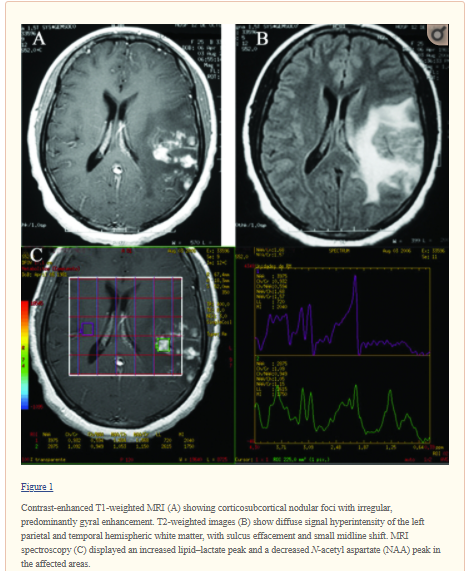
Liu H, Lim CC, Feng X, Yao Z, Chen Y, Sun H, Chen X. MRI in cerebral schistosomiasis: characteristic nodular enhancement in 33 patients. AJR Am J Roentgenol. 2008 Aug;191(2):582-8. doi: 10.2214/AJR.07.3139. PMID: 18647936.

Abstract

Objective: The purpose of our study was to describe the characteristic MRI appearance of cerebral infection with Schistosoma japonicum.

Conclusion: Cerebral infection with S. japonicum can cause a characteristic MRI pattern of a large mass comprising multiple intensely enhancing nodules, sometimes with areas of linear enhancement. The typical appearance may be useful for diagnosis in endemic regions and may potentially be useful in cases imported into countries in which the disease is not endemic.


Courtesy of a colleague

Thursday, March 25, 2021

Prolonged epileptic discharges predict seizure recurrence in JME

Turco F, Bonanni E, Milano C, Pizzanelli C, Steinwurzel C, Morganti R, Fornai F, Maestri M, Siciliano G, Giorgi FS. Prolonged epileptic discharges predict seizure recurrence in JME: Insights from prolonged ambulatory EEG. Epilepsia. 2021 Mar 18. doi: 10.1111/epi.16875. Epub ahead of print. PMID: 33735449.

Abstract

Objective: Markers of seizure recurrence are needed to personalize antiseizure medication (ASM) therapy. In the clinical practice, EEG features are considered to be related to the risk of seizure recurrence for genetic generalized epilepsies (GGE). However, to our knowledge, there are no studies analyzing systematically specific EEG features as indices of ASM efficacy in GGE. In this study, we aimed at identifying EEG indicators of ASM responsiveness in Juvenile Myoclonic Epilepsy (JME), which, among GGE, is characterized by specific electroclinical features. 

Methods: We compared the features of prolonged ambulatory EEG (paEEG, 22 h of recording) of JME patients experiencing seizure recurrence within a year ("cases") after EEG recording, with those of patients with sustained seizure freedom for at least 1 year after EEG ("controls"). We included only EEG recordings of patients who had maintained the same ASM regimen (dosage and type) throughout the whole time period from the EEG recording up to the outcome events (which was seizure recurrence for the "cases", or 1-year seizure freedom for "controls"). As predictors, we evaluated the total number, frequency, mean and maximum duration of epileptiform discharges (EDs) and spike density (i.e. total EDs duration/artifact-free EEG duration) recorded during the paEEG. The same indexes were assessed also in standard EEG (stEEG), including activation methods. 

Results: Both the maximum length and the mean duration of EDs recorded during paEEG significantly differed between cases and controls; when combined in a binary logistic regression model, the maximum length of EDs emerged as the only valid predictor. A cut-off of EDs duration of 2.68 seconds discriminated between cases and controls with a 100% specificity and a 93% sensitivity. The same indexes collected during stEEG lacked both specificity and sensitivity. 

Significance: The occurrence of prolonged EDs in EEG recording might represent an indicator of antiepileptic drug failure in JME patients.

Courtesy of:  https://www.mdlinx.com/journal-summary/prolonged-epileptic-discharges-predict-seizure-recurrence-in-jme-insights-from-prolonged-ambulatory/1PsinsUSGLh3qpxsEnNkP1


Elevated serum IL-10 is associated with severity of neonatal encephalopathy and adverse early childhood outcomes

Pang R, Mujuni BM, Martinello KA, Webb EL, Nalwoga A, Ssekyewa J, Musoke M, Kurinczuk JJ, Sewegaba M, Cowan FM, Cose S, Nakakeeto M, Elliott AM, Sebire NJ, Klein N, Robertson NJ, Tann CJ. Elevated serum IL-10 is associated with severity of neonatal encephalopathy and adverse early childhood outcomes. Pediatr Res. 2021 Mar 5. doi: 10.1038/s41390-021-01438-1. Epub ahead of print. PMID: 33674741.

Abstract

Background: Neonatal encephalopathy (NE) contributes substantially to child mortality and disability globally. We compared cytokine profiles in term Ugandan neonates with and without NE, with and without perinatal infection or inflammation and identified biomarkers predicting neonatal and early childhood outcomes. 

Methods: In this exploratory biomarker study, serum IL-1α, IL-6, IL-8, IL-10, TNFα, and VEGF (<12 h) were compared between NE and non-NE infants with and without perinatal infection/inflammation. Neonatal (severity of NE, mortality) and early childhood (death or neurodevelopmental impairment to 2.5 years) outcomes were assessed. Predictors of outcomes were explored with multivariable linear and logistic regression and receiver-operating characteristic analyses. 

Results: Cytokine assays on 159 NE and 157 non-NE infants were performed; data on early childhood outcomes were available for 150 and 129, respectively. NE infants had higher IL-10 (p < 0.001), higher IL-6 (p < 0.017), and lower VEGF (p < 0.001) levels. Moderate and severe NE was associated with higher IL-10 levels compared to non-NE infants (p < 0.001). Elevated IL-1α was associated with perinatal infection/inflammation (p = 0.013). Among NE infants, IL-10 predicted neonatal mortality (p = 0.01) and adverse early childhood outcome (adjusted OR 2.28, 95% CI 1.35-3.86, p = 0.002). 

Conclusions: Our findings support a potential role for IL-10 as a biomarker for adverse outcomes after neonatal encephalopathy. 

Impact: Neonatal encephalopathy is a common cause of child death and disability globally. Inflammatory cytokines are potential biomarkers of encephalopathy severity and outcome. In this Ugandan health facility-based cohort, neonatal encephalopathy was associated with elevated serum IL-10 and IL-6, and reduced VEGF at birth. Elevated serum IL-10 within 12 h after birth predicted severity of neonatal encephalopathy, neonatal mortality, and adverse early childhood developmental outcomes, independent of perinatal infection or inflammation, and provides evidence to the contribution of the inflammatory processes. Our findings support a role for IL-10 as a biomarker for adverse outcomes after neonatal encephalopathy in a sub-Saharan African cohort.

Courtesy of: https://www.mdlinx.com/journal-summary/elevated-serum-il-10-is-associated-with-severity-of-neonatal-encephalopathy-and-adverse-early/37md3Foi5S2uaJeJlo7GvR

Tuesday, March 23, 2021

Pandemic and social skills

DAYTON, OH—Unable to determine the root cause of the child’s complete emotional ineptitude, local parents Josh and Lindsey Stecher told reporters Tuesday it was unclear if their toddler Aiden’s social skills had been damaged by his isolation during the pandemic or if he was simply taking after Dad. “Sure, the reason he gets so nervous and freezes up in social situations could be because he’s spent so much time at home this past year, but he may also just be following in his father’s footsteps,” said Lindsey, explaining that the 1-year-old’s shy awkwardness and inability to make friends seemed “awfully familiar” and might have nothing at all to do with the fact that he had spent months staring at an iPad screen instead of playing with kids his own age. “On the one hand, Aiden is crippled with anxiety when he has to be around anyone besides me and Josh. On the other, it’s not really any different than what happens when I bring my husband to a party. I’m sure the quarantines take their toll, but so does being the child of a 38-year-old man who still hasn’t learned to maintain eye contact during a conversation and can’t get through an ordinary interaction like meeting a new neighbor without feeling embarrassed.” Regardless of what may have impaired his social development, the Stechers said they had resigned themselves to the fact that their son would grow up to be weird and painfully introverted just like his dad.

https://local.theonion.com/parents-can-t-tell-if-pandemic-inhibited-toddler-s-soci-1846514322

Courtesy of a colleague

Thursday, March 18, 2021

Team Hoyt

Dick Hoyt, who inspired thousands of runners, fathers and disabled athletes by pushing his son, Rick, in a wheelchair in dozens of Boston Marathons and hundreds of other races, has died, a member of the family said Wednesday.

He was 80.

Dick passed away quietly in his sleep at his Holland, Massachusetts, home on Wednesday morning, Russ Hoyt, another of his sons, told The Associated Press.

"He had an ongoing heart condition that he had been struggling with for years and it just got the better of him," Russ Hoyt said.

Russ and his other brother, Rob, broke the news to Rick.

"He's sad, as we all are, but he's OK," Russ said. "You could see it in him, it was like someone hit him."

Dick Hoyt first pushed his son, who is quadriplegic and has cerebral palsy, in the Boston Marathon in 1980. Dick and Rick, in a specialized wheelchair, completed 32 Boston Marathons together, until Dick, citing health issues, retired in 2014. He had planned on retiring after the 2013 race, but the father and son never finished because of that year's finish line bombing, so they came back one more time.

The Boston Athletic Association, which runs the marathon, called Hoyt a legend.

"The B.A.A. is tremendously saddened to learn of the passing of Boston Marathon icon Dick Hoyt," the organization said in a statement. "Dick personified what it meant to a be a Boston Marathoner, showing determination, passion, and love every Patriots Day for more than three decades. He was not only a fan favorite who inspired thousands, but also a loyal friend and father who took pride in spending quality time with his son Rick while running from Hopkinton to Boston."

Dick served as Grand Marshal of the 2015 race in recognition of his accomplishments.


And even though his father stopped, Rick did not. Bryan Lyons, a dentist, took over pushing Rick starting in 2015 until his unexpected death last June at age 50.

The 1980 Boston Marathon was not the Hoyts' first road race.

In 1977, Rick told his father that he wanted to participate in a benefit run for a lacrosse player who had been paralyzed. They finished next to last, but that was just the start.

"Dad, when I’m running, it feels like I’m not disabled," Rick told his father after that first race, according to the website of Team Hoyt, the charity the family established to help disabled athletes.

They participated in more than 1,000 races, including duathlons and triathlons and in 1992 even completed a run and bike across the U.S., covering 3,735 miles (6,010 kilometers) in 45 days, according to the website.

In 2013, a statue of father and son was erected in front of a school in Hopkinton, near the Boston Marathon's starting line.

"I know it's a cliche, but I want people to know that I thought my father was a hero, not just because he pushed Rick in the marathon, but because he was a great father to all of us you could talk to about anything," he said. "He inspired people to look at all their children as equals no matter their disability"

https://www.foxnews.com/sports/dick-hoyt-who-pushed-son-boston-marathons-dies
______________________________________________________________________________

The Early Years

Rick was born in 1962 to Dick and Judy Hoyt. As a result of oxygen deprivation to Rick’s brain at the time of his birth, Rick was diagnosed as a spastic quadriplegic with cerebral palsy. Dick and Judy were advised to institutionalize Rick because there was no chance of him recovering, and little hope for Rick to live a “normal” life. This was just the beginning of Dick and Judy’s quest for Rick’s inclusion in community, sports, education and one day, the workplace.

Dick and Judy soon realized that though Rick couldn’t walk or speak; he was quite astute and his eyes would follow them around the room. They fought to integrate Rick into the public school system, pushing administrators to see beyond Rick’s physical limitations. Dick and Judy would take Rick sledding and swimming, and even taught him the alphabet and basic words, like any other child. After providing concrete evidence of Rick’s intellect and ability to learn like everyone else, Dick and Judy needed to find a way to help Rick communicate for himself.

With $5,000 in 1972 and a skilled group of engineers at Tufts University, an interactive computer was built for Rick. This computer consisted of a cursor being used to highlight every letter of the alphabet. Once the letter Rick wanted was highlighted, he was able to select it by just a simple tap with his head against a head piece attached to his wheelchair. When the computer was originally first brought home, Rick surprised everyone with his first words. Instead of saying, “Hi, Mom,” or “Hi, Dad,” Rick’s first “spoken” words were: “Go, Bruins!” The Boston Bruins were in the Stanley Cup finals that season. It was clear from that moment on, that Rick loved sports and followed the game just like anyone else.

In 1975, at the age of 13, Rick was finally admitted into public school. After high school, Rick attended Boston University, and he graduated with a degree in Special Education in 1993. Dick retired in 1995 as a Lt. Colonel from the Air National Guard, after serving his country for 37 years.

The Beginning of Team Hoyt

In the spring of 1977, Rick told his father that he wanted to participate in a 5-mile benefit run for a Lacrosse player who had been paralyzed in an accident. Far from being a long-distance runner, Dick agreed to push Rick in his wheelchair and they finished all 5 miles, coming in next to last. That night, Rick told his father, “Dad, when I’m running, it feels like I’m not handicapped.”

This realization was just the beginning of what would become over 1,000 races completed, including marathons, duathlons and triathlons (6 of them being Ironman competitions). Also adding to their list of achievements, Dick and Rick biked and ran across the U.S. in 1992, completing a full 3,735 miles in 45 days.

In a triathlon, Dick will pull Rick in a boat with a bungee cord attached to a vest around his waist and to the front of the boat for the swimming stage. For the biking stage, Rick will ride a special two-seater bicycle, and then Dick will push Rick in his custom made running chair (for the running stage).

Rick was once asked, if he could give his father one thing, what would it be? Rick responded, “The thing I’d most like is for my dad to sit in the chair and I would push him for once.”

The 2009 Boston Marathon was officially Team Hoyt’s 1000th race. Rick always says if it comes down to doing one race a year he would like it to be the Boston Marathon: his favorite race.

2013 was going to be Dick and Rick's last Boston Marathon together, but they were not able to finish due to the bombings. They vowed to be back in 2014 to finish "Boston Strong" with all the other runners, which they did; stopping many times along the 26.2 distance to take photos and shake hands of the many well wishers, and finishing with several of the runners from their Hoyt Foundation Boston Marathon team.

Dick and Rick will continue to do shorter distances races. Neither Dick or Rick are ready to retire yet.


http://www.teamhoyt.com/About-Team-Hoyt.html

Wednesday, March 17, 2021

Real-world study shows steroid switching effective for dystrophinopathies

In a real-world study, individuals with Duchenne muscular dystrophy (DMD) and Becker muscular dystrophy (BMD) switched from being treated with prednisone to deflazacort (Emflaza; PTC Therapeutics, South Plainfield, NJ) to delay disease progression. For those who switched who had a Clinical Global Impression (CGI) scale scores recorded, most had disease progression improve or stabilize during steroid treatment, with a shift toward improvement after switching.

"We're encouraged by the clinical benefit exhibited by patients taking Emflaza in this real-world analysis," said Stuart W. Peltz, PhD, chief executive officer, PTC Therapeutics. "This data supports the results that we saw in patients on corticosteroids in the placebo arms of multiple Duchenne clinical trials and what we have heard from the Duchenne community. We are committed to providing access to clinically differentiated treatments for patients with high unmet need."

The real-world chart review was conducted with collected data from 92 male participants with DMD or BMD who switched from prednisone to deflazacort. The most common motivationsfor switching to deflazacort was desire to slow disease progression, tolerability, or caregiver or patient request. Of the 92 participants, 62 had DMD with an average age of 6.2 years during the switch and 30 had BMD with an average age of 20.1 years. The average treatment duration was 3.3 years for prednisone and 6 months for deflazacort prior to the chart extraction. Switching was reported as "very" or "somewhat" effective at addressing primary reasons in 95% of participants with DMD and 90% of participants with BMD.

"Results from the real-world chart review presented today support the potential of Emflaza to alter the natural history of Duchenne muscular dystrophy, demonstrating its capability to slow progression of the disease and improve benefit-risk," said Dr. Susan Apkon, investigator, chief Pediatric Rehabilitation, vice-chair Department of Physical Medicine and Rehabilitation, Fischahs chair in Pediatric Rehabilitation, Children's Hospital of Colorado. "We believe that Emflaza will continue to provide DMD patients a safe and effective treatment option."

The most common adverse events found when treated with prednisone and deflazacort included weight gain, Cushingoid appearance, increased appetite, central obesity, and fluid retention.

These data were presented at the 2021 Muscular Dystrophy Association (MDA) Virtual Clinical and Scientific Conference.

https://practicalneurology.com/news/real-world-study-shows-steroid-switching-effective-for-dystrophinopathies

Monday, March 15, 2021

Clinical features, treatment strategies, and outcomes in hospitalized children with immune-mediated encephalopathies.

McGetrick ME, Varughese NA, Miles DK, Wang CX, McCreary M, Monson NL, Greenberg BM MD. Clinical Features, Treatment Strategies, and Outcomes in Hospitalized Children With Immune-Mediated Encephalopathies. Pediatr Neurol. 2021 Mar;116:20-26. doi: 10.1016/j.pediatrneurol.2020.11.014. Epub 2020 Nov 28. PMID: 33388545.

Abstract

Background: Autoimmune encephalitis (AE) and acute disseminated encephalomyelitis (ADEM) are immune-mediated brain conditions that can cause substantial neurological sequalae. Data describing the clinical characteristics, treatments, and neurological outcomes for these conditions are needed.

Methods: This is a single-center retrospective review of children diagnosed with AE or ADEM over a nine-year period with discharge outcomes measured by the Modified Rankin Score.

Results: Seventy-five patients (23 with ADEM and 52 with AE) were identified. Patients with ADEM had a higher percentage of abnormal magnetic resonance imaging findings (100% vs 60.8%; P < 0.001) and a shorter time from symptom onset to diagnosis (6 vs 14 days; P = 0.024). Oligoclonal bands and serum and cerebrospinal fluid inflammatory indices were higher in patients with AE. Nearly all patients received corticosteroids followed by plasmapheresis or intravenous immunoglobulin, and treatment strategies did not differ significantly between groups. Second-line immune therapies were commonly used in patients with AE. Finally, patients with AE had trends toward longer hospital lengths of stay (21 vs 13 days) and a higher percentage of neurological disability at hospital discharge (59.6% vs 34.8%).

Conclusions: Although patients with ADEM and AE may have similar presenting symptoms, we found significant differences in the frequency of imaging findings, symptom duration, and laboratory and cerebrospinal fluid profiles, which can assist in distinguishing between the diagnoses. Patients in both groups were treated with a combination of immunomodulating therapies, and neurological disability  was common at hospital discharge.      ______________________________________________________________________

The most common forms of immune-related pediatric encephalitis are acute disseminated encephalomyelitis (ADEM) and autoimmune encephalitis (AE). Although these are two distinct disorders, explains UTSW pediatric critical care fellow Molly E. McGetrick, M.D., their presentation -- including disorientation and other signs of altered mental status, seizures, or motor and sensory abnormalities -- is largely the same in children, hindering an accurate diagnosis. In addition, the rarity of AE and ADEM makes amassing data to help distinguish these conditions more difficult.

"Despite their similarities in presentation, patients with AE tend to have a more prolonged and protracted condition that requires more therapies than those with ADEM," McGetrick explains. "Being able to definitively distinguish between these conditions could help doctors guide patients and their families on what to expect."...

McGetrick notes that currently ADEM and AE patients are given similar therapies, including corticosteroids to reduce the body's inflammatory reaction to autoantibodies, intravenous immunoglobulins to bind and neutralize pathologic autoantibodies, or plasmapheresis to remove autoantibodies from the body over a series of sessions. Many times, symptoms for both conditions will resolve with these treatments, but they can take longer for AE and recur in some individuals. The more researchers can learn about the distinguishing characteristics of these conditions, she says, the more they may be able to target specific treatments for each condition, improving the outlook for these patients.

"One of the biggest take-home messages from this study is that we still have a lot to learn about these conditions," McGetrick says. "The more we know, the brighter the future will ultimately be for these patients."

https://www.sciencedaily.com/releases/2021/02/210223121627.htm

Wednesday, March 10, 2021

Amelioration of levetiracetam- induced behavioral side effects by pyridoxine

Adel Mahmoud, Sadia Tabassum, Shoaa Al Enazi, Nahed Lubbad, Ali Al Wadei, Ali Al Otaibi, Lamya Jad and Ruba Benini. Amelioration of Levetiracetam- induced behavioral side effects by Pyridoxine-Randomized double blinded controlled study. Article in Press: Pediatric Neurology.

Abstract

Background

Levetiracetam is a relatively new generation antiseizure drug approved for the treatment of focal and generalized seizures. Despite its favorable side effect profile and minimal drug-drug interactions, neuropsychiatric side effects are reported in up to 13% of children. A few case series have suggested that supplementation of pyridoxine may mitigate these side effects, but controlled trials are lacking. To address this issue, a randomized interventional study was carried out in a pediatric tertiary hospital to qualify and quantify the potential beneficial effect of pyridoxine in attenuating the neuropsychiatric side effects of Levetiracetam in children. 

Methods

A total of 105 children with epilepsy on levetiracetam (as a monotherapy or an adjunct) who showed behavioral symptoms coinciding with the start of levetiracetam, were included. Patients randomly and blindly received either a therapeutic (pyridoxine group, 46/105, 44%) or a homeopathic dose of pyridoxine (placebo, 59/105, 56%). A 30-item behavioral checklist was used to qualify and quantify the behavioral side effects at baseline and at different time points following initiation of treatment. 

Results

Both Placebo and pyridoxine groups experienced a statistical reduction in behavioral scores as compared to baseline. Our study indicated that although there was a placebo effect, the improvement in neuropsychiatric symptoms was more prominent in children who received therapeutic doses of pyridoxine. 

Conclusion

This data provides clinicians with pertinent evidence-based information that suggests that a trial of pyridoxine in patients who experience behavioral side effects due to the use of levetiracetam may avoid unnecessary change of antiseizure medications.

KEY POINTS

• Epilepsy is a common neurological disorder in children in pediatrics worldwide.

• Levetiracetam is an antiseizure drug that has broad spectrum of action with good efficacy, low side effect profile and minimal drug-drug interactions.

• Behavioral side effects are the commonest with the use of levetiracetam.

• Pyridoxine is claimed to ameliorate behavioral side effects due to levetiracetam.

• This prospective randomized controlled trial provides evidence for the efficacy of pyridoxine in mitigating the behavioral side effects of levetiracetam.

• No adverse side effects were reported with pyridoxine use in the recommended doses.


Tuesday, March 9, 2021

Remote electrical neuromodulation for acute treatment of migraine in adolescents

Hershey, A.D., Lin, T., Gruper, Y., Harris, D., Ironi, A., Berk, T., Szperka, C.L. and Berenson, F. (2021), Remote electrical neuromodulation for acute treatment of migraine in adolescents. Headache: The Journal of Head and Face Pain, 61: 310-317. https://doi.org/10.1111/head.14042

Abstract

Objectives

Migraine is a common disabling neurological disorder. Current acute treatments for migraine in adolescents are mostly pharmacological and may have limited effectiveness, can cause side effects, and may lead to medication overuse. There is an unmet need for effective and welltolerated treatments. Remote electrical neuromodulation (REN) is a novel acute treatment of migraine that stimulates upper arm peripheral nerves to induce conditioned pain modulation (CPM)—an endogenous analgesic mechanism. The REN device (Nerivio®, Theranica BioElectronics Ltd., Israel) is a FDAauthorized device for acute treatment of migraine in adults. This study assessed the efficacy and safety of REN in adolescents with migraine.

Design and Methods

This was an openlabel, singlearm, multicenter study in adolescents (ages 12–17 years) with migraine. Participants underwent a 4week runin phase. Eligible participants continued to an 8week treatment phase with the device. Pain severity, associated symptoms, and functional disability were recorded at treatment initiation, and 2 and 24 hours posttreatment. The primary endpoints of this study were related to the safety and tolerability of REN. The secondary endpoints were related to device efficacy and included the proportion of participants who achieved pain relief at 2 hours posttreatment and the proportion of participants who achieved pain freedom at 2 hours. The presented results reflect an interim analysis with subsequent stopping of the rest of the study.

Results

Sixty participants were enrolled for the study; of these, 14 failed to meet the runin criteria and 1 was lost to followup. Fortyfive participants performed at least one treatment, of which 39 participants completed a test treatment with REN. One devicerelated adverse event (2%) was reported in which a temporary feeling of pain in the arm was felt. Pain relief and painfree at 2 hours were achieved by 71% (28/39) and 35% (14/39) participants, respectively. At 2 hours, 69% (23/33) participants experienced improvement in functional ability.

Conclusions

REN may offer a safe and effective nonpharmacological alternative for acute treatment in adolescents.

 Courtesy of:  https://www.mdlinx.com/journal-summary/remote-electrical-neuromodulation-for-acute-treatment-of-migraine-in-adolescents/4mq821S5QdC7nEjyPzQHw7

Monday, March 8, 2021

Infanticide vs. inherited cardiac arrhythmias

Brohus M, Arsov T, Wallace DA, Jensen HH, Nyegaard M, Crotti L, Adamski M, Zhang Y, Field MA, Athanasopoulos V, Baró I, Ribeiro de Oliveira-Mendes BB, Redon R, Charpentier F, Raju H, DiSilvestre D, Wei J, Wang R, Rafehi H, Kaspi A, Bahlo M, Dick IE, Chen SRW, Cook MC, Vinuesa CG, Overgaard MT, Schwartz PJ. Infanticide vs. inherited cardiac arrhythmias. Europace. 2020 Nov 17:euaa272. doi: 10.1093/europace/euaa272. Epub ahead of print. PMID: 33200177.

Abstract

Aims: In 2003, an Australian woman was convicted by a jury of smothering and killing her four children over a 10-year period. Each child died suddenly and unexpectedly during a sleep period, at ages ranging from 19 days to 18 months. In 2019 we were asked to investigate if a genetic cause could explain the deaths, as part of an inquiry into the mother's convictions.

Methods and results: Whole genomes or exomes of the mother and her four children were sequenced. Functional analysis of a novel CALM2 variant was performed by measuring Ca2+-binding affinity, interaction with calcium channels and channel function. We found two children had a novel calmodulin variant (CALM2 G114R) that was inherited maternally. Three genes (CALM1-3) encode identical calmodulin proteins. A variant in the corresponding residue of CALM3 (G114W) was recently reported in a child who died suddenly at age 4 and a sibling who suffered a cardiac arrest at age 5. We show that CALM2 G114R impairs calmodulin's ability to bind calcium and regulate two pivotal calcium channels (CaV1.2 and RyR2) involved in cardiac excitation contraction coupling. The deleterious effects of G114R are similar to those produced by G114W and N98S, which are considered arrhythmogenic and cause sudden cardiac death in children.

Conclusion: A novel functional calmodulin variant (G114R) predicted to cause idiopathic ventricular fibrillation, catecholaminergic polymorphic ventricular tachycardia, or mild long QT syndrome was present in two children. A fatal arrhythmic event may have been triggered by their intercurrent infections. Thus, calmodulinopathy emerges as a reasonable explanation for a natural cause of their deaths.                                  _______________________________________________________________________

The tabloids in Australia called Kathleen Folbigg a murderer of innocent babies — the nation’s “worst female serial killer.” In 2003, a court sentenced her to 40 years in prison for smothering her four children before each had turned 2.

But all along, Ms. Folbigg has insisted that she is innocent, and that her children were all victims of Sudden Infant Death Syndrome.

Now, 90 leading scientists say they’re convinced she is right. New genetic evidence, the scientists say, suggests that the children died from natural causes, and they are demanding that she be pardoned.

In a petition sent to the governor of New South Wales last week, the group of scientists, which includes two Nobel laureates, called for Ms. Folbigg’s immediate release and an end to the “miscarriage of justice.”

The very public challenge sets up a tense standoff between some of the world’s top medical minds and a criminal court system that rarely overturns convictions. It’s a story of judges putting more weight on the ambiguous musings of a mother’s diary than on rare genetic mutations, and of scientists who are determined to make the legal system respect cutting-edge expertise.

Caught in the middle is Ms. Folbigg, who is now 53. More than 30 years after her first child’s death, her story has not changed, and she maintains that she will be vindicated.

A Troubled Mother and Her Children

Ms. Folbigg’s life has been troubled almost since the moment she was born.

She was just 18 months old when her father, Thomas Britton, murdered her mother in 1968. His wife had walked out on them over a money dispute. He stabbed her on a public footpath in Sydney in a drunken rage.

Roughly 28 years later, Ms. Folbigg wrote in her diary: “Obviously, I am my father’s daughter.”

By that point, in 1996, she had married a miner, Craig Folbigg, had moved to a working-class suburb, Newcastle, a coal capital north of Sydney, and had lost three of her children.

Ms. Folbigg’s first child, Caleb, died on Feb. 20, 1989, at 19 days of age. His death was classified by doctors as Sudden Infant Death Syndrome, or SIDS.

The next child, Patrick, died nearly two years later, at 8 months. He was blind and had epilepsy and choked to death, according to his death certificate.

A baby girl, Sarah, died on Aug. 30, 1993, at 10 months old, and her death was also classified as SIDS. Ms. Folbigg’s last child, Laura, died in March 1999 at 18 months old, with the cause initially listed as “undetermined.”

The deaths seemed at first to be simple, horrific tragedy. But Ms. Folbigg’s husband turned her in to the police after reading one of her diary entries. It said Sarah had left “with a bit of help.”

Ms. Folbigg told the authorities that what she wrote had simply captured the angst and despair of young motherhood and that “a bit of help” referred to her hope that God had taken her baby home.

At her trial, the doctor who had ruled Laura’s death as undetermined, Allan Cala, testified that he had never seen a case of four children dying in the same family. He was admitted as an expert witness, and though he did not present independent data, prosecutors relied on his account to argue that lightning strikes and flying pigs were more likely than four babies dying so young in the same family over a span of 10 years.

“There has never, ever been in the history of medicine any case like this,” one prosecutor said in closing arguments. “It is not a reasonable doubt, it is preposterous.”

The jury agreed. Ms. Folbigg, 35 at the time, was found guilty of the murders of Patrick, Sarah and Laura and the manslaughter of Caleb. She collapsed into tears as the verdicts were read.

The Science That Could Set Her Free

But there was never any medical evidence of smothering, the scientists say — that was one hole in the case. It’s the first thing mentioned in their pardon petition for Ms. Folbigg.

None of the children, they go on to say, were healthy when they died. Laura, the last to die, had been sick with a respiratory infection, and an autopsy later found an inflamed heart.

With those hints in mind, her lawyers asked geneticists to examine the case, searching for a mutation that might explain the family’s experience.

Carola Vinuesa, an immunologist from the Australian National University in Canberra, and another doctor, Todor Arsov, visited Kathleen in prison on Oct. 8, 2018, and received consent to sequence her genome. They both found that Ms. Folbigg had a rare mutation in what’s known as the CALM2 gene.

The genetic defect essentially creates heart arrhythmias that can cause cardiac arrest and sudden death in infancy and childhood.

Only about 75 people in the world are known to have the mutation, Professor Vinuesa said, including some parents without symptoms. But children died in at least 20 of those cases, and in many others, they suffered cardiac arrest.

That was especially true when there were triggers driving up adrenaline — and one known trigger is pseudoephedrine, a drug Laura was taking when she died.

Using blood and tissue samples from all four children, taken shortly after they were born, Professor Vinuesa and Dr. Arsov found that Sarah and Laura both had the same mutation as their mother.

By that point, Ms. Folbigg’s lawyers, who had already exhausted formal appeals, managed to secure a formal inquiry into the case. Professor Vinuesa submitted a lengthy report in December 2018.

But there were signs of resistance. Dr. Cala re-emerged, telling the judge that by the time Laura’s body arrived, after three deaths, you “have to have in the back of your mind, is there something else going on in relation to possible trauma?”

Bob Moles, a law professor at Flinders University, said that the admission of such statements showed a major flaw in Australian justice.

“One of the main problems we have is a willingness of courts to admit scientific evidence that is not really scientific,” he said.

Sensing that the evidence was not being taken seriously, Professor Vinuesa wrote to Peter Schwartz, a world-leading genetic researcher in Milan. He wrote back and said he had been studying a family in the United States with the same mutation, including two children who died from heart attacks.

He sent a letter to the inquiry with his findings. In July 2019, the judge reached a decision. He said that he had considered the scientific evidence but that he had found Ms. Folbigg’s diary quite compelling — and that he had no reasonable doubt about her guilt.

Refusing to Give Up

Frustrated but more determined, the scientists’ network gradually expanded.

Several of the people involved, including Dr. Arsov, submitted their findings to an international peer-reviewed journal. The paper was published in November.

Further research into Caleb’s and Patrick’s genomes has revealed that they had a separate rare genetic variant, which in studies with mice has been linked to early lethal epileptic fits.

In all, 90 eminent scientists have agreed that the medical evidence proves Ms. Folbigg’s innocence. The signatories to the pardon petition include Dr. Schwartz; John Shine, president of the Australian Academy of Science; and Elizabeth Blackburn, a 2009 Nobel laureate in medicine who teaches at the University of California, San Francisco.

“We would feel exhilarated for Kathleen if she is pardoned,” Professor Vinuesa said. “It would send a very strong message that science needs to be taken seriously by the legal system.”

by Damien Cave

https://www.nytimes.com/2021/03/08/world/australia/kathleen-folbigg-child-murder-genetics.html?referringSource=articleShare

Courtesy of a colleague
Friday, March 5, 2021

Recovery of consciousness and functional outcome in moderate and severe traumatic brain injury

Kowalski RG, Hammond FM, Weintraub AH, et al. Recovery of Consciousness and Functional Outcome in Moderate and Severe Traumatic Brain Injury. JAMA Neurol. Published online March 01, 2021. doi:10.1001/jamaneurol.2021.0084

Key Points

Question  What are the trajectory of and factors associated with recovery of consciousness in patients with a disorder of consciousness (DOC) after traumatic brain injury (TBI)? 

Findings  In this cohort study of 17 470 patients with TBI, 57% of patients experienced initial loss of consciousness, which persisted after acute care treatment in 12% of patients. However, 98% of these patients recovered consciousness by the end of subsequent inpatient rehabilitation, and their trajectory of functional improvement mirrored that of patients with TBI who did not lose consciousness. 

Meaning  Results of this study indicated that most individuals who became comatose after moderate or severe TBI recovered consciousness in the short term and almost half of them regained functional independence, suggesting that caution is warranted in early decisions to withdraw or withhold treatment in patients with TBI and a DOC.

Abstract

Importance  Traumatic brain injury (TBI) leads to 2.9 million visits to US emergency departments annually and frequently involves a disorder of consciousness (DOC). Early treatment, including withdrawal of life-sustaining therapies and rehabilitation, is often predicated on the assumed worse outcome of disrupted consciousness. 

Objective  To quantify the loss of consciousness, factors associated with recovery, and return to functional independence in a 31-year sample of patients with moderate or severe brain trauma. 

Design, Setting, and Participants  This cohort study analyzed patients with TBI who were enrolled in the Traumatic Brain Injury Model Systems National Database, a prospective, multiyear, longitudinal database. Patients were survivors of moderate or severe TBI who were discharged from acute hospitalization and admitted to inpatient rehabilitation from January 4, 1989, to June 19, 2019, at 1 of 23 inpatient rehabilitation centers that participated in the Traumatic Brain Injury Model Systems program. Follow-up for the study was through completion of inpatient rehabilitation. 

Exposures  Traumatic brain injury. 

Main Outcomes and Measures  Outcome measures were Glasgow Coma Scale in the emergency department, Disability Rating Scale, posttraumatic amnesia, and Functional Independence Measure. Patient-related data included demographic characteristics, injury cause, and brain computed tomography findings. 

Results  The 17 470 patients with TBI analyzed in this study had a median (interquartile range [IQR]) age at injury of 39 (25-56) years and included 12 854 male individuals (74%). Of these patients, 7547 (57%) experienced initial loss of consciousness, which persisted to rehabilitation in 2058 patients (12%). Those with persisting DOC were younger; had more high-velocity injuries; had intracranial mass effect, intraventricular hemorrhage, and subcortical contusion; and had longer acute care than patients without DOC. Eighty-two percent (n = 1674) of comatose patients recovered consciousness during inpatient rehabilitation. In a multivariable analysis, the factors associated with consciousness recovery were absence of intraventricular hemorrhage (adjusted odds ratio [OR], 0.678; 95% CI, 0.532-0.863; P = .002) and intracranial mass effect (adjusted OR, 0.759; 95% CI, 0.595-0.968; P = .03). Functional improvement (change in total functional independence score from admission to discharge) was +43 for patients with DOC and +37 for those without DOC (P = .002), and 803 of 2013 patients with DOC (40%) became partially or fully independent. Younger age, male sex, and absence of intraventricular hemorrhage, intracranial mass effect, and subcortical contusion were associated with better functional outcome. Findings were consistent across the 3 decades of the database. 

Conclusions and Relevance  This study found that DOC occurred initially in most patients with TBI and persisted in some patients after rehabilitation, but most patients with persisting DOC recovered consciousness during rehabilitation. This recovery trajectory may inform acute and rehabilitation treatment decisions and suggests caution is warranted in consideration of withdrawing or withholding care in patients with TBI and DOC. ________________________________________________________________________

Most patients who become comatose after experiencing moderate or severe traumatic brain injury (TBI) recover consciousness in the short term — and nearly half regain functional independence, new research suggests. 

The study, which included more than 17,000 patients who were hospitalized with moderate and severe TBI over three decades, showed that even when they remained unconscious at the end of their initial acute hospital care and were admitted for subsequent inpatient rehabilitation, 82% recovered consciousness by rehab completion. 

"The results of our study, we think, show that caution is warranted in making decisions to withdraw or hold care in patients with these serious brain injuries," lead author Robert G. Kowalski, MBBCh, Department of Neurology, University of Colorado School of Medicine, Aurora, told Medscape Medical News. 

"A meaningful recovery is possible, even when loss of consciousness occurs after the brain injury," he added…

Historically, the prognosis of recovery for patients who have prolonged unconsciousness or disorders of consciousness (DOC) "has been perceived to be poor, with little hope for a return to independence," he said. 

Therefore, in a significant proportion of cases, decisions are made to withdraw or withhold life-sustaining therapies, and the patients subsequently die. "This in turn contributes to the perception of poor prognosis in severe TBI — a so-called 'self-fulfilling prophecy,' " Kowalski noted. 

The investigators evaluated the trajectory of, and factors associated with, recovery of consciousness and functional ability in patients with a DOC after moderate to severe TBI, focusing on the acute stage of emergent and critical care and subsequent inpatient rehabilitation. 

"We chose this period of care, including the initial hospitalization and subsequent inpatient rehabilitation, because this is the time window during which treating medical teams and families make critical decisions that may prolong life and affect longer-term outcome for these patients, and help determine how successfully they are able to return to independent living," Kowalski said…

However, 1674 comatose patients (82%) recovered consciousness (ability to follow commands) by the end of inpatient rehabilitation (median rehabilitation stay, 33 days). In addition, their trajectory of functional improvement mirrored that of patients with TBI who did not lose consciousness.

The investigators also observed the absence of specific signs of neuroanatomic injury on brain imaging, typically brain CT in the acute phase of treatment, including blood in the ventricles of the brain and severe midline shift of cerebral structures. This absence portends better prospects for recovery of consciousness and functional ability for these patients, the researchers note…

In an accompanying editorial, Jennifer Kim, MD, PhD, and Kevin Sheth, MD, Division of Neurocritical Care, Yale School of Medicine, New Haven, Connecticut, note that the study "further challenges our potential toward overly nihilistic notions of who may, or may not, ultimately recover consciousness long term" by showing that a large proportion of patients with persistent DOC recover during acute rehabilitation. 

"Other studies that followed up patients long term (not restricted to the inpatient rehabilitation period) corroborate the observation that recovery in TBI can occur 6 to 12 months after injury," they write.

The current study used one of the largest cohorts of patients with TBI available to assess recovery in the rehabilitation setting, and the "remarkable rate of recovery should give pause to practitioners who counsel families about potential recovery of DoC," write Kim and Sheth.

"If there are no concerning radiographic features, then practitioners should communicate the potential for delayed DoC recovery," they add. 

Echoing the investigators, the editorialists write that this study "adds to the TBI literature cautioning against withdrawal of life-sustaining therapy even when faced with prolonged DoC during hospitalization because there remains significant potential for recovery." 

"Defining both good and poor prognostic risk factors is critical to portending recovery. Future work must refine biomarker identification and use in patients with DoC to improve physician prognostication and avoid self-fulfilling prophecy," they conclude.

https://www.medscape.com/viewarticle/946867


Thursday, March 4, 2021

Single brain death examination Is equivalent to dual brain death examinations

Varelas, P.N., Rehman, M., Abdelhak, T. et al. Single Brain Death Examination Is Equivalent to Dual Brain Death Examinations. Neurocrit Care 15, 547–553 (2011). https://doi.org/10.1007/s12028-011-9561-4 

Abstract

Background

Although the new Practice Parameters for brain death support a single examination, there is paucity of data comparing its impact to dual brain death (DBD) examinations. 

Methods

We reviewed all brain deaths in our hospital over a 39-month period and compared the optional single brain death (SBD) exam requiring an apnea and a mandatory confirmatory blood flow test to the DBD for organ function at the time of death, rate of donation, and cost. 

Results

Thirty-six patients had a SBD and 59 DBD exams, without any of them regaining neurological functioning. There was no difference in serum electrolytes (except for higher Na+ and Cl− in the SBD group), blood urea nitrogen, creatinine, blood gases, incidence of diabetes insipidus, apnea completion, consent for donation, and organs recovered and transplanted. During the second BD exam, 35% of patients with DBD were on higher dose of vasopressors, but had lower systolic blood pressure (P = 0.046). For DBD patients, the mean interval between the two exams was 14.4 h, which contributed to a higher cost of $43,707.67 compared to SBD. There was a trend for increased consent rates (adjusted for age, race, and type of exam) when patients were declared by the neurointensivist service following a strict family approach protocol (P = 0.06). 

Conclusion

SBD exam is easier, faster to perform, with no brain function recovery and leads to similar donation rates, equivalent or better organ function status at the time of BD and lower cost than conventional DBD exams.

More sunflowers

James R. Barnett, Bradley M. Fleming, Samarth P. Doshi, Jason Freedman, Nicholas R. Ambrosio, Kennedy R. Geenen, Patricia L. Bruno, Elizabeth A. Thiele. Understanding Sunflower syndrome: Results of an online questionnaire. Epilepsy & Behavior, Volume 117, 2021. https://doi.org/10.1016/j.yebeh.2021.107856.

Abstract

Objective

To characterize the clinical phenotype, treatments, and impact on quality of life of Sunflower syndrome. 

Methods

A 138-question survey was created focusing on seizure description, disease course, treatment history, medical history, family history, and aspects of quality of life of individuals with Sunflower syndrome. The survey was administered to individuals with Sunflower syndrome who experience hand waving episodes (HWE) and/or their caregivers via Research Electronic Data Capture (REDCap). 

Results

Sixty-eight responses were included in analysis. Seventy-one% of respondents were female. The mean age of participants was 13.6years, with 84% of respondents under the age of 18. The average age of onset of HWE was 6.7years. HWE frequency varied from a few episodes per week to multiple episodes per hour. Sixty-two% of participants experienced other seizure types. Participants had been on an average of 1.9 anti-seizure medications with varying efficacy. Other methods to reduce HWE included wearing a hat or sunglasses, hand holding, using special tinted lenses, and avoiding the sun and bright lights. Sixty-nine% of participants reported anxiety or depression related to their epilepsy, and 65% said their HWE affected their social life. 

Significance

Sunflower syndrome is a highly stereotyped, refractory epilepsy which significantly impacts the lives of affected individuals. It remains underrecognized and poorly understood. These results characterize Sunflower syndrome in a large population of affected individuals and provides a basis for future research to better understand the epilepsy and improve clinical care.

See: https://childnervoussystem.blogspot.com/2021/01/sunflower-syndrome.html

Courtesy of a patient's parent 

The wilderness of rare genetic diseases and the parents navigating it

Anne and Jerry Van Wyk didn’t notice anything unusual about their son, Reid, in the days after his birth in June 2001. But on his 10th day, when Anne Van Wyk tried to wake her newborn, she thought his breathing was irregular.

Jerry Van Wyk observed something was off, too. They rushed their son to a clinic near their home in Mankato, Minn., and then to a hospital emergency room, where doctors discovered Reid had an enlarged liver. He was airlifted to Minneapolis, where he underwent a battery of tests over the next four days.

Jerry Van Wyk observed something was off, too. They rushed their son to a clinic near their home in Mankato, Minn., and then to a hospital emergency room, where doctors discovered Reid had an enlarged liver. He was airlifted to Minneapolis, where he underwent a battery of tests over the next four days.

Doctors told the couple that Reid’s arteries were turning to stone. Calcium was building up, blocking the flow of blood, and straining his heart. There were fewer than 200 documented cases worldwide of generalized arterial calcification of infancy, or GACI. Most were not surviving past their first year. “They sent us home without much hope at all,” Anne Van Wyk said.

About a month later Reid died. He was seven weeks.

At the time, there was no test available to examine the genetic cause of GACI, and almost no information about the rare genetic disease online, the Van Wyks said. Over the next decade, the couple experienced six miscarriages and another death linked to the condition. They had three healthy children during that time, but still felt cursed and alone. “How could this be happening with us?” Jerry Van Wyk asked. 

Rare diseases are quite common, conversely. In the United States, the National Institutes of Health defines a rare disease as any condition affecting fewer than 200,000 people. But the 7,000 or so known rare diseases affect as many as 10 percent of the U.S. population. About 80 percent are genetic and about half appear in children, many of whom don’t survive into adulthood. 

Rare DiseasesMeet four families who succeeded in promoting research on uncommon diseases, but in very different ways.

Shortly after learning that their child has a rare disease, parents often feel alone as they navigate a sea of acronyms, a maze of hospital corridors and piles of unpaid bills. Some will start to lose faith in their doctors and the medical system to provide for their child’s particular needs. But treatment options for children have gotten better in recent years — in part, as a result of parents’ grit. 

The parents, also called “rare parents,” have discovered power in numbers. They’ve learned how to connect with other families experiencing what they are going through, and then join forces to raise money and support research that could change their child’s life. 

“When I was practicing clinical genetics, it was limited to diagnosis and prognosis,” said Jim Wilson, M.D., Ph.D., the director of the gene therapy program and the orphan disease center at the University of Pennsylvania’s School of Medicine. “Now, in a limited number of diseases, there are potential treatments, if not cures.” 

A confirmed diagnosis may take time.

Diagnosis represents the first step on this rare disease journey. Sometimes doctors will notice something off about the child during a newborn screening, and a genetic test will identify a known mutation in the DNA. But not all conditions are so quickly detected, and it can take several years for parents to get a confirmed diagnosis. 

About half of all children never get that far, according to Marshall Summar, M.D., the director of the Rare Disease Institute at Children’s National Hospital in Washington, D.C. “When you sequence someone’s DNA, you are going to find a lot of changes,” Dr. Summar said. “Figuring out which change might be the one that is causing it is a tremendous challenge.” 

Genetic counselors warn parents beforehand that they may not get a definitive answer as to what condition their child could have. They may have to check back each year. Dr. Summar estimates that between five and 10 new rare diseases are described in the scientific literature every week, making it challenging for the medical field to keep up. 

Meanwhile, the realization that a child may have a debilitating, lifelong condition weighs heavily. “Some parents, particularly mothers, blame themselves,” said Lemuel Pelentsov, Ph.D., a nurse who studies the needs of rare disease families at the University of South Australia, in Adelaide. In a 2016 study by Dr. Pelentsov and his colleagues, about 40 percent of the 300 rare parents surveyed reported being treated for depression and an equal number for anxiety. “One of the things they do to combat that,” he said, “is get very invested in the child’s disease.” 

Families will find support in each other.

When parents reach out to other parents, they are not simply looking for emotional support or advice. They are rebuilding a social life, one that will revolve around their child’s disease. Many rare diseases have their own support groups. Global Genes is an umbrella group that supports 600 disease-specific foundations, as well as parents of children whose diseases are so rare they have no foundation. 

“We encourage folks to work together,” said Kimberly Haugstad, the organization’s executive director whose son has a rare form of hemophilia, a condition in which the blood doesn’t clot normally. “The parent is going to come from such different places in their own walk of life.” 

Some parents will start nonprofits or fund research.

Each year, Global Genes hosts a “Rare Boot Camp” to mentor and teach parents how to set up a nonprofit, create patient registries and fund research. After attending the boot camp, the Van Wyks and other parents founded GACI Global, an organization that connects families affected by GACI, along with medical professionals. 

Adding to the unknowns, parents must figure out if they want to create a support group, raise awareness or fund research. Rather than setting up a small nonprofit specifically for a rare disease, they may decide to establish a research fund with a pre-existing organization. 

Parents may influence the making of new drugs.

If the research is far enough along, families also seek ways to accelerate cures or therapies, forging relationships with scientists and companies. 

For common diseases, like heart disease or diabetes, pharmaceutical companies compete with each other to get patients to ask their doctors for their drugs. With rare diseases the situation is essentially reversed, with parents courting researchers and drugmakers to get even a single drug on the market for their children. To do this, parents compile detailed records of symptoms and create polished presentations to persuade them the disease is a good investment.

 Every parent brings their own set of skills. In October 2012, Maria Picone’s daughter was born with Prader-Willi syndrome, which causes severe intellectual disability. After joining a Facebook group called “Living Well with Prader-Willi Syndrome,” Picone learned from other parents that children were also passing out frequently, a symptom that wasn’t widely studied or properly treated. 

Picone and her husband, who both had a background in software development, started a company called TREND Community that turns such data-rich Facebook discussions into what she calls “Community Voice Reports.” Those reports can then be shared with companies and regulators, like the U.S. Food and Drug Administration. 

Dr. Wilson said his 360-person team at the University of Pennsylvania’s School of Medicine not only runs clinical trials on potential therapies, but the Orphan Disease Center, which is part of the School of Medicine, also distributes up to $5 million in grants each year to other research teams in the rare disease space. “The question is how do you select what disease or what disease groups to work with?” Wilson asked. “I wish I could tell you there is a magic formula.” 

While the Orphan Drug Act in the United States has helped get new drugs for rare diseases on the market, its exclusivity provision, critics say, has kept drug prices high. Some parents and organizations have kept a financial stake in technologies coming out of the research they fund, or founded their own companies to develop drugs. These moves give them more control in the drug development process and the potential to ensure that the drugs are affordable to those who need them. 

The Foundation for Angelman Syndrome Therapeutics, for example, an all voluntary organization of families and professionals, hopes to commercialize a drug candidate for the neurological disease. Clinical trials began in January. That accomplishment “speaks volumes to the determination of some parents,” said the organization’s founder, Paula Evans. 

As for the Van Wyks, they ended up having the large family they always wanted. In late 2012, however, the couple learned that their youngest daughter, Natalie, tested positive for GACI while in utero. The ultrasound showed that calcification was rapidly progressing. The couple reached out to a specialist in Germany that they had been in contact with since Reid’s death. They wanted to know if there was anything they could do for Natalie. 

He sent the couple a 1992 report of a child who had been successfully treated with an osteoporosis drug while still in the womb, at Children’s Hospital of Philadelphia. It was an experimental treatment and would require close monitoring. Anne Van Wyk moved to Philadelphia to undergo treatment, and could be monitored over the course of the pregnancy. 

In 2013, Natalie was born. Although she has hearing loss and will have to take a phosphorus supplement and calcium-boosting medicine for the rest of her life, she will be starting first grade in the fall near their new home in Fort Worth, Tex. 

Six out of seven GACI babies have now survived in the last eight years, thanks to the prenatal treatment given at that hospital, according to Michael A. Levine, M.D., director of the hospital’s Center for Bone Health. Dr. Levine plans to publish the results in a medical journal, but in the meantime, the treatment’s existence and potential has spread through word-of-mouth and been shared on the GACI Global website. “We are glad to be that glimmer of hope in a dark space,” Anne Van Wyk said.

https://www.nytimes.com/2020/07/07/parenting/rare-genetic-diseases-children.html?referringSource=articleShare

Courtesy of a colleague


Increased mortality in very young children with traumatic brain injury due to abuse

 Christina M. Theodorou, Miriam Nuño, Kaeli J. Yamashiro, Erin G. Brown. Increased Mortality in Very Young Children with Traumatic Brain Injury Due to Abuse: A Nationwide Analysis of 10,965 Patients. Published:February 24, 2021DOI:https://doi.org/10.1016/j.jpedsurg.2021.02.044 

Abstract

Background

: Traumatic brain injury (TBI) is the leading cause of death and disability in young children; however, the impact of mechanism on outcomes has not been fully evaluated. We hypothesized that children with TBI due to abuse would have a higher mortality than children with accidental TBI due to motor vehicle collisions (MVC).

Methods

: We performed a retrospective review of the National Kids’ Inpatient (KID) hospitalizations database of children <2 years old with TBI due to abuse or MVC (2000-2016). The primary outcome was mortality. Secondary outcomes were length of stay (LOS) and hospital charges. We investigated predictors of mortality with multivariable regression.

Results

: Of 10,965 children with TBI, 65.2% were due to abuse. Overall mortality was 9.8% (n=1074). Abused children had longer LOS (5.7 vs 1.6 days, p<0.0001) and higher hospital charges ($34,314 vs $19,360, p<0.0001) than children with TBI due to MVC. The odds of mortality were 42% higher in children with abusive head trauma (OR 1.42, 95% CI 1.10-1.83, p=0.007) compared to MVCs after adjusting for age, race, sex, neurosurgical intervention, injury severity, and insurance.

Conclusion

: Children with abusive traumatic brain injury have increased risk of mortality, longer LOS, and higher hospital charges compared to children with TBI due to motor vehicle collision after adjusting for relevant confounders. Resources must be directed at prevention and early identification of abuse.

Courtesy of:  https://www.mdlinx.com/journal-summary/increased-mortality-in-very-young-children-with-traumatic-brain-injury-due-to-abuse-a-nationwide/1xRCvg28CgjPuqCcBrUS9a

Transparency update 3

A state appeals court Wednesday upheld a decision requiring CNN to provide copies of emails and text messages as the network fights a lawsuit filed by a physician who contends he was defamed in news reports about a pediatric heart-surgery program at St. Mary’s Medical Center in West Palm Beach. 

A three-judge panel of the 4th District Court of Appeal rejected arguments that CNN should be shielded from turning over the documents because of a state law that protects journalists from having to provide information related to sources. The ruling said the law offers “qualified privilege” and agreed that physician Michael Black was entitled to see copies of communications between CNN journalists and Kelly Robinson, a source in the coverage of the hospital. 

Black, who was the head of the pediatric heart-surgery program, filed the defamation lawsuit in 2016 against CNN, individual journalists --- including star anchor Anderson Cooper --- and Robinson. CNN published and broadcast reports in June 2015, saying, in part, that the surgery program had a high infant-mortality rate. The hospital disputed the reports, and Black alleges in the lawsuit that he was the target of “false and defamatory” statements.

A Palm Beach County circuit judge in May ordered CNN to turn over the emails and text messages, and the appeals court said Wednesday that the judge “found a compelling interest for disclosure because of the unique circumstances of the case.” Among those circumstances was the need for Black to prove malice in the lawsuit and the fact that Robinson, who is not a journalist, had deleted her copies of the communications. 

“While the journalist privilege must be protected, it is a qualified privilege,” said the six-page ruling, written appeals-court Judge Martha Warner and joined by Chief Judge Spencer Levine and Judge Burton Conner. “(A section of state law) provides the procedure to overcome that privilege when the facts and circumstances of a case require it. The trial court concluded that respondent (Black) overcame the privilege in this case. The trial court did not depart from any clearly established principle of law.” 

The CNN reports about the St. Mary’s program drew widespread attention, with Black’s lawsuit and the appeals court ruling describing Kennedy as a “key source” of information for the network. The Palm Beach Post reported in 2015 that Kennedy’s son had heart surgery at the rival Nicklaus Children’s Hospital in Miami and that she was highly critical of St. Mary’s and Black. 

The appeals court found that the emails and text messages between CNN journalists and Robinson were relevant to the case. 

“We reject CNN’s argument that the communications are irrelevant given its claim that Robinson was not the source of any specific defamatory statement contained in CNN’s reporting,” the ruling said. “Even if Robinson was not the source of the calculations of mortality rates, it is clear that there were communications between Robinson and CNN about those rates and the respondent. Also, Robinson remained a key source and connection in the case. Thus, those communications would show what CNN ‘knew or did not know at the time of publication,’ both on behalf of CNN and also Robinson, who is also a defendant in the case.”

https://cbs12.com/news/local/cnn-required-to-hand-over-emails-in-west-palm-beach-doctor-defamation-case

See:  https://childnervoussystem.blogspot.com/2015/06/transparency.html

https://childnervoussystem.blogspot.com/2017/03/transparency-update.html


Tuesday, March 2, 2021

Janus kinase inhibition in the Aicardi-Goutières syndrome

Vanderver A, Adang L, Gavazzi F, McDonald K, Helman G, Frank DB, Jaffe N, Yum SW, Collins A, Keller SR, Lebon P, Meritet JF, Rhee J, Takanohashi A, Armangue T, Ulrick N, Sherbini O, Koh J, Peer K, Besnier C, Scher C, Boyle K, Dubbs H, Kramer-Golinkoff J, Pizzino A, Woidill S, Shults J. Janus Kinase Inhibition in the Aicardi-Goutières Syndrome. N Engl J Med. 2020 Sep 3;383(10):986-989. doi: 10.1056/NEJMc2001362. PMID: 32877590; PMCID: PMC7495410. 

The Aicardi–Goutières syndrome is a genetic interferonopathy that is associated with severe disability and death. Most children with this syndrome are unable to walk or talk and have multisystemic complications, including skin inflammation (figure in article). Janus kinase (JAK) inhibitors may be effective in blocking interferon activation in patients with the Aicardi–Goutières syndrome. 

We conducted an open-label study of a single-center, expanded-access program (ClinicalTrials.gov numbers, NCT01724580. opens in new tab and NCT03047369. opens in new tab; see the protocol, available with the full text of this letter at NEJM.org) involving 35 patients with genetically confirmed Aicardi–Goutières syndrome. The patients received baricitinib, an oral JAK1 and JAK2 inhibitor. Eli Lilly provided the medication for the study and provided support for safety monitoring. 

The median age at the onset of symptoms was 6.0 months, and the median age at the initiation of baricitinib was 2.9 years (range, 0.2 to 21.8). The patients participated in the study for a minimum of 12.0 months (range, 11.8 to 43.8). The dose of baricitinib, which ranged from 0.1 to 0.6 mg per kilogram of body weight per day, administered in 2 to 4 dosing increments per day, was determined according to the patient’s renal function, age, and symptoms.

JAK inhibitors are associated with infection, anemia, lymphopenia, and thrombosis. Two patients died during the study: one who had been receiving glucocorticoids for a decade, including a 7-year period before the study, had Aicardi–Goutières syndrome–related multisystem organ failure with an opportunistic infection, and the other had Aicardi–Goutières syndrome–related pulmonary hypertension with thrombotic microangiopathy. Clinical variables were measured with laboratory tests and graded according to Common Terminology Criteria for Adverse Events, version 5.0, in which grades range from 1 to 5, with higher grades indicating more severe adverse events. Among these variables, only thrombocytosis increased in severity during the study (mean change in grade from baseline, 0.30; 95% confidence interval [CI], 0.04 to 0.60). 

A biomarker for interferon signaling, the interferon-signaling gene-expression score, indicated a response to treatment (figure in article). This score is calculated from the expression of six interferon-signaling genes normalized to housekeeping genes. The six-gene interferon signature is the sum of the median z scores of these genes. The interferon signature is positive (i.e., type 1 interferon signaling is high) if it is at least 1.96 (>98th percentile) on a one-tailed test (see the Supplemental Methods section in the Supplementary Appendix, available at NEJM.org). 

The parents of the patients used diaries to record the children’s Aicardi–Goutières syndrome–related symptoms, including neurologic disability, crying, sleep disturbances, irritability, seizures, fever, and skin inflammation of the trunk, arms, and legs (see the Supplemental Methods section and Table S5 in the Supplementary Appendix in article). The diary score included a total of 24 possible points, assigned every day, with daily scores averaged across the time period before each visit (1 month after the initiation of baricitinib and then every 3 months). Scores obtained during the study ranged from 0 to 10.6, with higher scores indicating more severe symptoms. The skin subscore included a total of 6 possible points, with higher scores indicating more severe symptoms. The overall diary scores improved within 1 month after the initiation of therapy (mean change in overall score, −0.7 diary points; 95% CI, −1.2 to −0.2) and were sustained (mean change in overall score, −1.4; 95% CI, −2.2 to −0.6) (figures in article). The percentage of patients with a lower (improved) skin subscore of only 0 or 1 increased from 66% (23 of 35 patients) to 83% (29 of 35 patients) from the baseline visit to the final visit (difference, 17 percentage points; 95% CI, 2 to 32). 

We evaluated the patients’ developmental histories from disease onset (median age since disease onset, 16.5 months; interquartile range, 7.4 to 41.5) to the clinical data cutoff date (figure in article). We extracted information on key milestones, including head control, sitting, rolling, assisted and independent ambulation, grasp, smiling, babbling, and the use of single words and word combinations. Before treatment, 26 of the 35 patients had stable or declining neurologic function, and 9 of the 35 patients gained one or two skills after disease onset. During the study, 20 patients met new milestones, and 12 patients gained two to seven new skills. This improvement was noted by 3 months (mean change in the number of milestones, 0.5; 95% CI, 0.2 to 0.9) and persisted (mean change, 1.3; 95% CI, 0.6 to 1.9). One patient lost skills during an acute illness while participating in the study. Children in a higher daily dose category of baricitinib met more milestones (mean increase, 1.3 milestones; 95% CI, 0.6 to 1.9) than those in a lower daily dose category according to weight-based calculations (figure in article). 

The primary risks associated with baricitinib among patients with the Aicardi–Goutières syndrome were thrombocytosis, leukopenia, and infection (see Section 4.1.2 in the Supplementary Appendix in article). Patients who are receiving baricitinib should be monitored closely, especially those with underlying thrombotic risk factors or those who are receiving systemic glucocorticoids or immunosuppressive regimens. Because this study drew from an international population, we anticipate that our findings will be generalizable. The measurement of neurologic improvement is complex, but our data suggest improvement in neurologic function, even in patients with severe and long-standing disease.