Monday, December 31, 2018

Inherited GPI deficiencies

Inspired by a patient

Nguyen TTM, Murakami Y, Wigby KM, Baratang NV, Rousseau J, St-Denis A, Rosenfeld JA, Laniewski SC, Jones J, Iglesias AD, Jones MC, Masser-Frye D, Scheuerle AE, Perry DL, Taft RJ, Le Deist F, Thompson M, Kinoshita T, Campeau PM. Mutations in PIGS, Encoding a GPI Transamidase, Cause a Neurological Syndrome Ranging from Fetal Akinesia to Epileptic Encephalopathy. Am J Hum Genet. 2018 Oct 4;103(4):602-611.

Abstract
Inherited GPI deficiencies (IGDs) are a subset of congenital disorders of glycosylation that are increasingly recognized as a result of advances in whole-exome sequencing (WES) and whole-genome sequencing (WGS). IGDs cause a series of overlapping phenotypes consisting of seizures, dysmorphic features, multiple congenital malformations, and severe intellectual disability. We present a study of six individuals from three unrelated families in which WES or WGS identified bi-allelic phosphatidylinositol glycan class S (PIGS) biosynthesis mutations. Phenotypes included severe global developmental delay, seizures (partly responding to pyridoxine), hypotonia, weakness, ataxia, and dysmorphic facial features. Two of them had compound-heterozygous variants c.108G>A (p.Trp36) and c.101T>C (p.Leu34Pro), and two siblings of another family were homozygous for a deletion and insertion leading to p.Thr439_Lys451delinsArgLeuLeu. The third family had two fetuses with multiple joint contractures consistent with fetal akinesia. They were compound heterozygous for c.923A>G (p.Glu308Gly) and c.468+1G>C, a splicing mutation. Flow-cytometry analyses demonstrated that the individuals with PIGS mutations show a GPI-AP deficiency profile. Expression of the p.Trp36 variant in PIGS-deficient HEK293 cells revealed only partial restoration of cell-surface GPI-APs. In terms of both biochemistry and phenotype, loss of function of PIGS shares features with PIGT deficiency and other IGDs. This study contributes to the understanding of the GPI-AP biosynthesis pathway by describing the consequences of PIGS disruption in humans and extending the family of IGDs.

Yang L, Peng J, Yin XM, Pang N, Chen C, Wu TH, Zou XM, Yin F. Homozygous PIGT Mutation Lead to Multiple Congenital Anomalies-Hypotonia Seizures Syndrome 3. Front Genet. 2018 May 8;9:153.

PIGT encodes a subunit of the glycosylphosphatidylinositol transamidase complex, which catalyzes the attachment of proteins to GPI-anchors. A homozygous PIGT variant c.550G>A (p. E184K) in a Chinese boy with multiple malformations, hypotonia, seizure and profound development delay was identified by panel sequencing. Pathogenicity of the variant was confirmed by flow cytometry. The expression of CD16 and CD24 of this proband reduced to 16.92 and 22.16% compare with normal control respectively while which of his parents and sister were normal. This mutation raised the mRNA level on the peripheral blood mono nuclear cells of this patient. This study expanded the variant spectrum of MCAHS3, and CD16 could be an effective marker to evaluate the pathogenicity of PIGT mutation.

Nguyen TTM, Murakami Y, Sheridan E, Ehresmann S, Rousseau J, St-Denis A, Chai G, Ajeawung NF, Fairbrother L, Reimschisel T, Bateman A, Berry-Kravis E, Xia F, Tardif J, Parry DA, Logan CV, Diggle C, Bennett CP, Hattingh L, Rosenfeld JA,  Perry MS, Parker MJ, Le Deist F, Zaki MS, Ignatius E, Isohanni P, Lönnqvist T, Carroll CJ, Johnson CA, Gleeson JG, Kinoshita T, Campeau PM. Mutations in GPAA1, Encoding a GPI Transamidase Complex Protein, Cause Developmental Delay, Epilepsy, Cerebellar Atrophy, and Osteopenia. Am J Hum Genet. 2017 Nov 2;101(5):856-865.

Abstract
Approximately one in every 200 mammalian proteins is anchored to the cell membrane through a glycosylphosphatidylinositol (GPI) anchor. These proteins play important roles notably in neurological development and function. To date, more than 20 genes have been implicated in the biogenesis of GPI-anchored proteins. GPAA1 (glycosylphosphatidylinositol anchor attachment 1) is an essential component of the transamidase complex along with PIGK, PIGS, PIGT, and PIGU (phosphatidylinositol-glycan biosynthesis classes K, S, T, and U, respectively). This complex orchestrates the attachment of the GPI anchor to the C terminus of precursor proteins in the endoplasmic reticulum. Here, we report bi-allelic mutations in GPAA1 in ten individuals from five families. Using whole-exome sequencing, we identified two frameshift mutations (c.981_993del [p.Gln327Hisfs102] and c.920delG [p.Gly307Alafs11]), one intronic splicing mutation (c.1164+5C>T), and six missense mutations (c.152C>T [p.Ser51Leu], c.160_161delinsAA [p.Ala54Asn], c.527G>C [p.Trp176Ser], c.869T>C [p.Leu290Pro], c.872T>C [p.Leu291Pro], and c.1165G>C [p.Ala389Pro]). Most individuals presented with global developmental delay, hypotonia, early-onset seizures, cerebellar atrophy, and osteopenia. The splicing mutation was found to decrease GPAA1 mRNA. Moreover, flow-cytometry analysis of five available individual samples showed that several GPI-anchored proteins had decreased cell-surface abundance in leukocytes (FLAER, CD16, and CD59) or fibroblasts (CD73 and CD109). Transduction of fibroblasts with a lentivirus encoding the wild-type protein partially rescued the deficiency of GPI-anchored proteins. These findings highlight the role of the transamidase complex in the development and function of the cerebellum and the skeletal system.

Sunday, December 30, 2018

KBG syndrome


Inspired by http://childnervoussystem.blogspot.com/2018/12/portrait-evoking-negative-emotions.html

Morel Swols D, Tekin M. KBG Syndrome. 2018 Mar 22. In: Adam MP, Ardinger HH, Pagon RA, Wallace SE, Bean LJH, Stephens K, Amemiya A, editors. GeneReviews® [Internet]. Seattle (WA): University of Washington, Seattle; 1993-2018. Available from http://www.ncbi.nlm.nih.gov/books/NBK487886/

Excerpt
CLINICAL CHARACTERISTICS:
KBG syndrome is typically characterized by macrodontia (especially of the upper central incisors), characteristic facial features (triangular face, brachycephaly, synophrys, widely spaced eyes, broad or bushy eyebrows, prominent ears, prominent nasal bridge, bulbous nose, anteverted nares, long philtrum, and thin vermilion of the upper lip), short stature, developmental delay / intellectual disability, and behavioral issues. Affected individuals may have feeding difficulties (particularly in infancy), skeletal anomalies (brachydactyly, large anterior fontanelle with delayed closure, scoliosis), hearing loss (conductive, mixed, and sensorineural), seizure disorder, and brain malformations. There is significant variability in the clinical findings, even between affected members of the same family.

DIAGNOSIS/TESTING:
The diagnosis of KBG syndrome is confirmed in a proband by detection of either a heterozygous pathogenic variant in ANKRD11 or deletion of 16q24.3 that includes ANKRD11.

MANAGEMENT:
Treatment of manifestations. Surgical corrections and/or speech therapy for palatal anomalies; nasogastric tube feeding in infants; pharmacologic treatment for gastroesophageal reflux disease; pressure-equalizing tubes and/or tonsillectomy/adenoidectomy for chronic otitis media; consideration of amplification for hearing loss; consideration of growth hormone therapy for short stature and medication to arrest puberty for premature pubertal development; standard treatment of seizure disorder, undescended testis in males, congenital heart defects, strabismus / refractive errors, and developmental disabilities. Surveillance. Routine monitoring of hearing, vision, growth, pubertal status (in prepubertal individuals), and cognitive development. Agents/circumstances to avoid. Ototoxic drugs should be avoided because of the risk for hearing loss. Pregnancy management. Pregnancy management should be tailored to the specific features in the affected woman. For example, involvement of a cardiologist and maternal fetal medicine physician for a pregnant woman with a history of a congenital heart defect; control of seizures during pregnancy for those with a seizure disorder.

GENETIC COUNSELING:
Recurrence risk for sibs of a proband with KBG syndrome depends on the genetic alteration: Deletion of 16q24.3 (~75% of reported pathogenic variants are de novo and the remainder are inherited in an autosomal dominant manner.) ANKRD11 sequence variants (~66% of reported pathogenic variants are de novo and the remainder are inherited in an autosomal dominant manner.) Prenatal testing and preimplantation genetic diagnosis are possible if the causative genetic alteration has been identified in an affected family member.

De Bernardi ML, Ivanovski I, Caraffi SG, Maini I, Street ME, Bayat A, Zollino M, Lepri FR, Gnazzo M, Errichiello E, Superti-Furga A, Garavelli L. Prominent and elongated coccyx, a new manifestation of KBG syndrome associated with novel mutation in ANKRD11. Am J Med Genet A. 2018 Sep;176(9):1991-1995.

Abstract
KBG syndrome is characterized by short stature, distinctive facial features, and developmental/cognitive delay and is caused by mutations in ANKRD11, one of the ankyrin repeat-containing cofactors. After the advent of whole exome sequencing, the number of clinical reports with KBG diagnosis has increased, leading to a revision of the phenotypic spectrum associated with this syndrome. Here, we report a female child showing clinical features of the KBG syndrome in addition to a caudal appendage at the coccyx with prominent skin fold and a peculiar calcaneus malformation. Exons and exon-intron junctions targeted resequencing of SH3PXD2B and MASP1 genes, known to be associated with prominent coccyx, gave negative outcome, whereas sequencing of ANKRD11 whose mutations matched the KBG phenotype of the proband showed a de novo heterozygous frameshift variant c.4528_4529delCC in exon 9 of ANKRD11. This report contributes to expand the knowledge of the clinical features of KBG syndrome and highlights the need to search for vertebral anomalies and suspect this condition in the presence of a prominent, elongated coccyx.

Behnert A, Auber B, Steinemann D, Frühwald MC, Huisinga C, Hussein K, Kratz C, Ripperger T. KBG syndrome patient due to 16q24.3 microdeletion presenting with a paratesticular rhabdoid tumor: Coincidence or cancer predisposition? Am J Med Genet A. 2018 Jun;176(6):1449-1454.


Abstract
KBG syndrome is a rare autosomal dominant disorder caused by constitutive haploinsufficiency of the ankyrin repeat domain-containing protein 11 (ANKRD11) being the result of either loss-of-function gene variants or 16q24.3 microdeletions. The syndrome is characterized by a variable clinical phenotype comprising a distinct facial gestalt and variable neurological involvement. ANKRD11 is frequently affected by loss of heterozygosity in cancer. It influences the ligand-dependent transcriptional activation of nuclear receptors and tumor suppressive function of tumor protein TP53. ANKRD11 thus serves as a candidate tumor suppressor gene and it has been speculated that its haploinsufficiency may lead to an increased cancer risk in KBG syndrome patients. While no systematic data are available, we report here on the second KBG syndrome patient who developed a malignancy. At 17 years of age, the patient was diagnosed with a left-sided paratesticular extrarenal malignant rhabdoid tumor. Genetic investigations identified a somatic truncating gene variant in SMARCB1, which was not present in the germline, and a constitutional de novo 16q24.3 microdeletion leading to a loss of the entire ANKRD11 locus. Thus, KBG syndrome was diagnosed, which was in line with the clinical phenotype of the patient. At present, no specific measures for cancer surveillance can be recommended for KBG syndrome patients. However, a systematic follow-up and inclusion of KBG syndrome patients in registries (e.g., those currently established for cancer prone syndromes) will provide empiric data to support or deny an increased cancer risk in KBG syndrome in the future.

Monteiro JP, Rijo D, Pereira R, Guerra M. Isolated tricuspid valve Staphylococcus lugdunensis endocarditis in patient with a KBG syndrome. Rev Port Cir Cardiotorac Vasc. 2018 Jan-Jun;25(1-2):91-93. Abstract in English, Portuguese

Both KBG syndrome (approximately 50 patients worldwide) and isolated tricuspid valve Staphylococcus lugdunensis endocarditis are very rare entities. The KBG syndrome is a multiple congenital anomaly characterized by short stature, distinctive craniofacial features, and neurologic/developmental/cognitive delay and is only associated to congenital heart defects in 9% of patients. Staphylococcus lugdunesis is an aggressive cause of infective endocarditis. Herein is described a case of a patient presenting both diseases, despite the absence of any known infection, congenital heart defect, heart device or any other entry port which could explain this scenario. The unusual findings in this young patient raised questions regarding the, as-yet unexplained, etiopathogenesis of the KBG syndrome, the possibility of it being related to this rare and concerning clinical presentation and the unclear and undefined management and surgical approach associated to right side endocarditis.

Thursday, December 27, 2018

Albuterol and congenital myasthenia


Liewluck T, Selcen D, Engel AG. Beneficial effects of albuterol in congenital endplate acetylcholinesterase deficiency and Dok-7 myasthenia. Muscle Nerve. 2011 Nov;44(5):789-94.

Abstract

INTRODUCTION:
Congenital myasthenic syndromes (CMS) are disabling but treatable disorders. Anticholinesterase therapy is effective in most of them, but is contraindicated in endplate (EP) acetylcholinesterase (AChE) deficiency, the slow-channel syndrome, Dok-7 myasthenia, and β(2) -laminin deficiency, and is not useful in CMS due to defects in muscle-specific kinase (MuSK), agrin, and plectin. EP AChE, Dok-7, and β(2)-laminin deficiencies respond favorably to ephedrine, but ephedrine can no longer be prescribed in the USA.

METHODS:
We used albuterol, another sympathomimetic agent, to treat 3 patients with EP AChE deficiency and 15 with Dok-7 myasthenia. Response to therapy was evaluated by a 9-point questionnaire pertaining to activities of daily life.

RESULTS:
Comparison of the pre- and posttreatment responses indicated a beneficial response to albuterol (P < 0.001) in both patient groups. The adverse effects of therapy were like those of ephedrine.

CONCLUSION:
Our observations should spur controlled, prospective clinical trials of albuterol in these as well as other CMS.

Farmakidis C, Pasnoor M, Barohn RJ, Dimachkie MM. Congenital Myasthenic Syndromes: a Clinical and Treatment Approach. Curr Treat Options Neurol. 2018 Jul 21;20(9):36.

Abstract

PURPOSE OF REVIEW:
Congenital myasthenia syndromes are clinically and genetically heterogeneous but treatable conditions. Careful selection of drug therapy is paramount as the same drug can be effective, ineffective, and even harmful in different congenital myasthenia syndromes. The purpose of this article is to review current treatment options for these conditions.

RECENT FINDINGS:
Next-generation sequencing has accelerated the discovery of new genes and facilitated the description of novel congenital myasthenic syndromes. Retrospective therapy data from these newly identified syndromes has provided additional insight on the management of these conditions. Cholinergic agents, β-adrenergic agonists, and open-channel blockers remain the principal treatment modalities, and their optimal use depends on an accurate genetic diagnosis and the timely clinical recognition of the disease. In particular, pyridostigmine, usually a first-line agent, should be avoided in DOK7, acetylcholinesterase deficiency, and slow-channel congenital myasthenic syndromes. Beta-adrenergic agonists have been recognized as a first-line agent for a number of congenital myasthenic syndromes, particularly DOK7 and acetylcholinesterase deficiency, whereas long-lived open-channel blockers of the acetylcholine receptor (AChR) ion channel are indicated for the slow-channel congenital myasthenic syndrome. Beta-adrenergic agonists additionally have an important adjunct treatment for congenital myasthenia syndrome due to glycosylation defects, fast channel syndrome, AChR deficiency, and choline acetyltransferase deficiency (ChaT) and therefore may be particularly important in the treatment of syndromes due to defects in motor endplate development and repair. Unlike in autoimmune myasthenia gravis, there is no role for immunotherapy in congenital myasthenic syndromes. If available, a genetic diagnosis should drive the choice for a first-line treatment agent between cholinergic agents, β-adrenergic agents, and open-channel blockers. Evaluation and supportive care at centers with experience in these rare syndromes likely are paramount in achieving optimal outcomes. Furthermore, gene discovery for congenital myasthenic syndromes has provided novel insights on the role of protein glycosylation, endplate maintenance and repair, and synaptic vesicle exocytosis in neuromuscular transmission. These insights may lead to new therapeutic strategies in both congenital and autoimmune myasthenic diseases in the future.

Evoli A, Alboini PE, Damato V, Iorio R, Provenzano C, Bartoccioni E, Marino M. Myasthenia gravis with antibodies to MuSK: an update. Ann N Y Acad Sci. 2018 Jan;1412(1):82-89.

Abstract
Myasthenia gravis with antibodies to the muscle-specific tyrosine kinase (MuSK+ MG) is a rare disease with distinctive pathogenic mechanisms and clinical features. An acute onset and predominant bulbar muscle weakness are very common and highly suggestive of the disease. On the other hand, a more indolent course, atypical ocular presentation, and signs of cholinergic hyperactivity may complicate the diagnosis. Though MuSK+ MG is still a severe disease, over the years we have observed a steady reduction in the rate of respiratory crisis and a significant improvement in the clinical outcome, both likely related to earlier diagnosis and timely treatment. Despite the improved management, MuSK+ MG patients tend to remain dependent on long-term immunosuppressive treatment and may develop permanent disabling weakness. In uncontrolled studies, B cell depletion with rituximab proved effective in most patients with refractory disease, inducing prolonged clinical responses associated with a sustained reduction of serum antibody levels. Promising results from experimental studies and case reports suggest that both 3,4-diaminopyridine and albuterol may be effective as symptomatic agents.

Ghazanfari N, Morsch M, Tse N, Reddel SW, Phillips WD. Effects of the ß2-adrenoceptor agonist, albuterol, in a mouse model of anti-MuSK myasthenia gravis. PLoS One. 2014 Feb 5;9(2):e87840. doi: 10.1371/journal.pone.0087840.

Abstract

The β2-adrenergic receptor agonist, albuterol, has been reported beneficial in treating several forms of congenital myasthenia. Here, for the first time, we examined the potential benefit of albuterol in a mouse model of anti-Muscle Specific Kinase (MuSK) myasthenia gravis. Mice received 15 daily injections of IgG from anti-MuSK positive patients, which resulted in whole-body weakness. At neuromuscular junctions in the tibialis anterior and diaphragm muscles the autoantibodies caused loss of postsynaptic acetylcholine receptors, and reduced the amplitudes of the endplate potential and spontaneous miniature endplate potential in the diaphragm muscle. Treatment with albuterol (8 mg/kg/day) during the two-week anti-MuSK injection series reduced the degree of weakness and weight loss, compared to vehicle-treated mice. However, the compound muscle action potential recorded from the gastrocnemius muscle displayed a decremental response in anti-MuSK-injected mice whether treated with albuterol or vehicle. Ongoing albuterol treatment did not increase endplate potential amplitudes compared to vehicle-treated mice nor did it prevent the loss of acetylcholine receptors from motor endplates. On the other hand, albuterol treatment significantly reduced the degree of fragmentation of endplate acetylcholine receptor clusters and increased the extent to which the remaining receptor clusters were covered by synaptophysin-stained nerve terminals. The results provide the first evidence that short-term albuterol treatment can ameliorate weakness in a robust mouse model of anti-MuSK myasthenia gravis. The results also demonstrate that it is possible for albuterol treatment to reduce whole-body weakness without necessarily reversing myasthenic impairment to the structure and function of the neuromuscular junction.

See:  https://childnervoussystem.blogspot.com/2015/05/i-am-appalled.html

Congenital myasthenia gravis


A 2-year-old girl with a history of celiac disease and eosinophilic esophagitis presented to the emergency department (ED) with 1 week of right-sided ptosis in the setting of isolated left-sided ptosis for 4 days during the prior week.

History. She had been seen initially by her primary care provider for evaluation of the symptoms, at which time significant dental caries had been noted, and the girl had been prescribed amoxicillin for a possible dental abscess. The left-sided ptosis then resolved; however, several days later, she developed similar symptoms on the right side with accompanying rhinorrhea. The ptosis appeared to worsen before sleep. A review of systems was negative for fever, eye drainage, lid swelling/erythema, abnormal gait, weakness, or generalized fatigue. Her medical history was significant for gross motor skill delay and expressive language delay.

Physical examination. In the ED, physical examination findings were notable for right-sided ptosis, with fatigability during prolonged upward gaze and abnormal left eye abduction causing dysconjugate gaze. Examination findings were otherwise unremarkable, with the exception of dental caries.

A pediatric neurologist was consulted in the setting of fatigable weakness, normal MRI findings, and diurnal variation of ptosis. Further testing included negative electromyography (EMG) results and negative test results for anti-acetylcholine receptor (AChR) and anti–muscle-specific tyrosine kinase (MuSK) antibodies.

Diagnostic tests.  A computed tomography scan of the head was obtained, the results of which revealed radiographic sinusitis without periorbital or orbital cellulitis. The results of cerebrospinal fluid studies were unremarkable. Given the persistence of symptoms, she was admitted to the hospital for further workup.

She was treated with intravenous ampicillin-sulbactam and intravenous amoxicillin-clavulanate for sinusitis without resolution of neurologic symptoms. Magnetic resonance imaging (MRI)/magnetic resonance venography with sedation was performed, the results of which demonstrated pansinusitis but most notably were negative for septic thromboembolism, mass lesion, optic neuritis, or demyelination. Specifically, the bilateral branches of the oculomotor nerve within the cavernous sinus appeared normal and did not explain the presence of ptosis. Furthermore, the optic chiasm and carotid arteries were without signs of compression to account for the fluctuating symptoms.

Treatment and outcome. She remained well appearing without other signs of illness, and thus she was transitioned to oral amoxicillin-clavulanate for sinusitis and was discharged with plans to follow up with the pediatric neurology clinic. Given her history of motor skill developmental delays and the fatigable nature of her ptosis, she was started on pyridostigmine, an acetylcholinesterase inhibitor, for presumed congenital myasthenia syndrome (CMS). She experienced clinical improvement on this medication, which was felt to be likely diagnostic of CMS in the absence of positive antibody test results. Pyridostigmine was slowly titrated to symptom management at follow-up visits with subsequent improvement.

Discussion. Causes of acquired ptosis include foreign bodies, trauma, allergic reactions, conjunctivitis, cellulitis, Horner syndrome, and intracranial mass. Rapidly progressive ptosis suggests a serious underlying problem that requires immediate evaluation. Conversely, congenital ptosis results from localized myogenic dysgenesis or denervation of the levator palpebrae superioris muscle via neurologic or neuromuscular dysfunction. Thus spinal muscular atrophy, muscular dystrophies, and brainstem anomalies should all be considered in the diagnosis.

CMS is one cause of congenital ptosis and is classified into 4 subtypes: presynaptic, postsynaptic (fast channel), postsynaptic (slow channel), and synaptic. It is inherited in an autosomal recessive fashion, apart from slow-channel CMS, which is autosomal dominant.  When CMS is suspected, a complete neurologic examination and ophthalmic evaluation should be performed, including visual acuity testing and refraction testing with dilation. Typically, patients with CMS will demonstrate fatigable weakness suggestive of neuromuscular junction disease.

Testing for anti-AChR and anti-MuSK antibodies should be performed, the results of which will be negative in this non–immune-mediated disorder. EMG studies generally show characteristics similar to disorders of synaptic transmission, although some phenotypes have unique features that can be seen on EMG.5 Obtaining an MRI may be helpful for ruling out other etiologies and for evaluation of fatty infiltration caused by mutations of the proteins involved in the glycosylation pathway. Many gene mutations have been implicated, and whole-gene sequencing may be performed. At this time, at least 11 mutations have been confirmed, predominantly of the postsynaptic type, such as RAPSN and CHRNE mutations.

Although CMS is a congenital syndrome, weakness can present variably from infancy to adulthood, with later presentations being classically milder. Response to treatments that are known to ameliorate neuromuscular transmission is a significant diagnostic and prognostic factor. Disease severity can range from early childhood death to minor morbidity.  It is important to recognize that, unlike myasthenia gravis, CMS is not an autoimmune disease and is unresponsive to immunosuppressive therapy. Furthermore, although juvenile myasthenia gravis (JMG) comprises the largest proportion of pediatric myasthenia cases, no cases of JMG have been documented in patients younger than 1 year of age; instead, this disease typically occurs in school-aged children.8

Early recognition and diagnosis is critical to optimize clinical management, anticipate complications, and provide appropriate genetic counseling. Genetic testing should be dictated by clinical features and presentation, since it requires DNA testing from both parents and can be costly for families.9 Our patient’s ocular findings, fatigable weakness, and age at presentation were fairly typical, as was her immediate response to pharmacologic intervention, despite normal EMG and MRI results, since findings may be intermittent in childhood.  Genetic testing in this patient’s case was unavailable as a result of insurance coverage limitations.

Treatment is largely supportive.5 However, expedited use of medication is crucial to preventing further morbidity, especially in patients with preexisting developmental delays. Most children benefit from acetylcholinesterase inhibitors (AChEIs) and/or a potassium-channel blocker (amifampridine). Additionally, prophylactic AChEIs may be used to prevent sudden episodes of apnea or respiratory insufficiency provoked by fever or infections.  Many cases of pediatric myasthenia are unrecognized or misdiagnosed given the subtleties of clinical findings. If familial genetic mutations are known, targeted testing can be used to identify asymptomatic newborns and infants to prevent acute respiratory failure and early death.

https://www.consultant360.com/articles/congenital-myasthenia-syndrome?hmpid=Z2JyZW5pbmdzdGFsbEBnaWxsZXR0ZWNoaWxkcmVucy5jb20=

Wednesday, December 26, 2018

Acute necrotizing encephalopathy


A basketball-obsessed boy is now wheelchair-bound and unable to verbally communicate after he was struck down by a brain damaging illness triggered by the flu. But despite the horrific illness, the Steph Curry-obsessed kid is making a remarkable bounce back.

Andre Carson, 4, spent 11 days on life support after he contracted the H1N1 flu virus, which triggered acute necrotizing encephalopathy (ANE).

The extremely rare condition causes lesions to develop in regions of the brain, which causes swelling, bleeding and eventually the death of the organ’s tissue.

According to the U.S. National Library of Medical Science, only 59 cases of ANE have been documented in scientific literature and one-third of patients with the illness do not survive.

Andre’s mom Kamareia Parrish, 29, was horrified to discover her child unresponsive and barely breathing hours after the family’s doctor had diagnosed him with a common cold on March 29, 2017.

At 2 a.m., Andre, of Fresno, Calif., was rushed by ambulance to a local hospital before he was transferred to Valley Children’s Hospital in Madera, where he was placed on life support. Although doctors diagnosed Andre with ANE and began treating the illness with steroids, they told Parrish he had suffered severe damage to his brain stem and the left side of his brain.

“Andre was a friendly, active and bubbly little boy," Parrish said. “He was already in preschool when he became sick. He was getting ready to start t-ball and soccer that weekend. That Monday his preschool called me and said he was feeling sick so I went and picked him up. He had a sore throat, a fever, diarrhea and he had chills so I brought him straight to the doctor."

"He suspected Andre was suffering from a cold so I treated him with Tylenol and I put him to bed," she said. “That night at about 2 a.m. I went to check on him and give him his sippy cup and he wasn’t responding to me. I tried to stand him up but he was just limp. He was barely breathing. I was so afraid I called an ambulance and when I saw it arrive I just ran outside with him straight away. They stabilized him and intubated him, but it was very serious."

"A CT scan revealed that there were lesions on his brain and they discovered he had the H1N1 flu virus," she said. “After he was transferred to the other hospital, the lesions just kept getting bigger and bigger so they started treating him with steroids. The steroids managed to reduce the swelling in his brain but his brain stem and the left side of his brain had a lot of damage.

“They diagnosed him with necrotizing encephalopathy, this was triggered but the H1N1 flu virus," she said. “It was my lowest moment. I was so angry with God. I was questioning my religion. I told my parents that if Andre died they might as well plan two funerals because I would die too. I had to believe he would live. He spent 11 days in the ICU in total."

“They extubated him that Sunday and when he started breathing on his own I was so hopeful," Parrish said. “But he opened his eyes and he wasn’t responding. While it was nice to see them open, he was like a vegetable.”

Andre spent two months in rehabilitation, where he began to voluntarily move his limbs and track movements with his eyes. The little boy returned home on June 13, 2017.

More than a year after falling ill, Andre can only communicate using an electronic device and is beginning to take his first steps using a walker.

The little boy receives his nutrition through a feeding tube and gets around using a power wheelchair.

Parrish was forced to give up her career to become Andre’s full-time carer and said she never knew the damage that could be caused by the flu.


“I was so surprised that this all stemmed from the flu," Parrish, a single mom, said. "Andre was vaccinated but it was for a different strain of the virus. I was really angry at myself at the beginning because I kept thinking, what if I had brought him to the hospital sooner but my doctor has told me there was no way I could have known."

"Vaccinations are so important. I work closely with an organization called the End- FLUenza Project, which was started by a mom who lost her daughter to a strain of the flu," she said. "I don’t know where Andre picked up the flu but maybe if someone had gotten the shot or taken precautions to prevent the spread of flu, he wouldn’t have gotten sick. I’m really paranoid about everything now. If I could keep Andre in a bubblesuit for his whole life I would."

"He’s pretty much in a wheelchair all the time but he is learning to walk using a walker and lets me know what he needs with his communications device," she said. "Our goals for the year are to speak two-word sentences, to start walking independently, to grow his vocab."

The mom has high hopes that Andre will keep progressing and says the fact that he is alive is a miracle.

"It’s been so hard to watch his friends from preschool move on and start school," she said. "But I have to stay positive because he’s lucky. Science says he shouldn't be here anymore. I know he will keep trying and fighting. I know he’s the same little boy inside but he’s just trapped in his body. I know this because he's still obsessed Steph Curry, just like he was before he got sick. He's still obsessed with basketball - that hasn't changed one bit."

To donate to the End-FLUenza project or learn more visit https://endfluenzaproject.org/

https://www.foxnews.com/health/boy-develops-rare-brain-eating-disease-after-contracting-flu?intcmp=ob_article_footer_text&intcmp=obnetwork


Tuesday, December 25, 2018

Deep brain stimulation in pediatric patients with GNAO1 associated severe hyperkinesia


Inspired by a patient

Koy A, Cirak S, Gonzalez V, Becker K, Roujeau T, Milesi C, Baleine J, Cambonie G, Boularan A, Greco F, Perrigault PF, Cances C, Dorison N, Doummar D, Roubertie A, Beroud C, Körber F, Stüve B, Waltz S, Mignot C, Nava C, Maarouf M, Coubes P, Cif L. Deep brain stimulation is effective in pediatric patients with GNAO1 associated severe hyperkinesia. J Neurol Sci. 2018 Aug 15;391:31-39.

Abstract

BACKGROUND:
Exacerbation of hyperkinesia is a life-threatening complication of dyskinetic movement disorders, which can lead to multi-organ failure and even to death. GNAO1 has been recently identified to be involved in the pathogenesis of early infantile epileptic encephalopathy and movement disorders. Patients with GNAO1 mutations can present with a severe, progressive hyperkinetic movement disorder with prolonged life-threatening exacerbations, which are refractory to most anti-dystonic medication.

OBJECTIVE:
The objective was to investigate the evolution of symptoms and the response to deep brain stimulation of the globus pallidus internus (GPi-DBS) in patients with different GNAO1 mutations.

METHODS:
We report six patients presenting with global motor retardation, reduced muscle tone and recurrent episodes of severe, life-threatening hyperkinesia with dystonia, choreoathetosis, and ballism since early childhood. Five of them underwent GPi-DBS.

RESULTS:
The genetic workup revealed mutations in GNAO1 for all six patients. These encompass a new splice site mutation (c.723+1G>T) in patient 1, a new missense mutation (c.610G>C; p.Gly204Arg) in patient 2, a heterozygous mutation (c.625>T; p.Arg209Cys) in patients 3 and 4, and a heterozygous mutation (c.709G>A; p.Glu237Lys) in patients 5 and 6. By intervention with GPi-DBS the severe paroxysmal hyperkinetic exacerbations could be stopped in five patients. One patient is still under evaluation for neuromodulation.

CONCLUSION:
In complex movement disorders of unsolved etiology clinical WES can rapidly streamline pathogenic genes. We identified two novel GNAO1 mutations. GPi-DBS can be an effective and life-saving treatment option for patients with GNAO1 mutations and has to be considered early.

Honey CM, Malhotra AK, Tarailo-Graovac M, van Karnebeek CDM, Horvath G, Sulistyanto A. GNAO1 Mutation-Induced Pediatric Dystonic Storm Rescue With Pallidal Deep Brain Stimulation. J Child Neurol. 2018 May;33(6):413-416.

Abstract

Dystonic storm or status dystonicus is a life-threatening hyperkinetic movement disorder with biochemical alterations due to the excessive muscle contractions. The medical management can require pediatric intensive care unit admission and a combination of medications while the underlying trigger is managed. Severe cases may require general anesthesia and paralytic agents with intubation and may relapse when these drugs are weaned. Deep brain stimulation of the globus pallidum has been reported to terminate dystonic storm in several pediatric cases. We present a 10-year-old boy with a de novo GNAO1 mutation-induced dystonic storm who required a 2-month pediatric intensive care unit admission and remained refractory to all medical treatments. Deep brain stimulation was performed under general anesthetic without complication. His dyskinetic movements stopped with initiation of stimulation. He was discharged from the pediatric intensive care unit after 4 days. We present prospectively evaluated changes in dystonia symptoms and quality of life for a patient with GNAO1 mutation treated with deep brain stimulation.

Feng H, Khalil S, Neubig RR, Sidiropoulos C. A mechanistic review on GNAO1-associated movement disorder. Neurobiol Dis. 2018 Aug;116:131-141.

Abstract

Mutations in the GNAO1 gene cause a complex constellation of neurological disorders including epilepsy, developmental delay, and movement disorders. GNAO1 encodes Gαo, the α subunit of Go, a member of the Gi/o family of heterotrimeric G protein signal transducers. Go is the most abundant membrane protein in the mammalian central nervous system and plays major roles in synaptic neurotransmission and neurodevelopment. GNAO1 mutations were first reported in early infantile epileptic encephalopathy 17 (EIEE17) but are also associated with a more common syndrome termed neurodevelopmental disorder with involuntary movements (NEDIM). Here we review a mechanistic model in which loss-of-function (LOF) GNAO1 alleles cause epilepsy and gain-of-function (GOF) alleles are primarily associated with movement disorders. We also develop a signaling framework related to cyclic AMP (cAMP), synaptic vesicle release, and neural development and discuss gene mutations perturbing those mechanisms in a range of genetic movement disorders. Finally, we analyze clinical reports of patients carrying GNAO1 mutations with respect to their symptom onset and discuss pharmacological/surgical treatments in the context of our mechanistic model.

Saturday, December 22, 2018

Portrait evoking negative emotions


Editor’s note: Two days after this article appeared, NIH director Francis S. Collins apologized to Beyond the Diagnosis about the portrait request described here. Collins said the NIH will display a series of portraits for its “Rare Disease Day and a future showing at the Clinical Center.”...

“Beyond the Diagnosis” is an exhibit of portraits of people — mostly children — with rare diseases that have been created by a small army of volunteer artists. More than that, it’s a presentation of the colorful souls within those seemingly imperfect bodies and a bright, uplifting, safe way to display them.

I was shocked that the show’s organizers were withdrawing it from a planned two-month stay at the National Institutes of Health. The NIH is renowned for its efforts in advancing medicine and improving the treatment of disease, including rare diseases. And it has hosted the exhibit before, as have the FDA, the Mayo Clinic, Harvard Medical School, and others.

What could have prompted this? According to the NIH Clinical Center, “the hospital art curator expressed to the exhibit organizer that one of the portraits in the collection of rare disease portraits may evoke negative emotions in patients.” I’ve seen the portrait the NIH wanted removed, and all I saw was a happy, vibrant young man.

My shock turned to anger. It was a visceral response, one I have, unfortunately, felt many times before.

When I was growing up in Utah, my sister was my best friend and greatest champion. As a youngster, I didn’t know she had special needs — she was just my sister. Once I realized that her life in school was made difficult by some of the other kids, I became her defender.

One day I arrived home from school as the phone rang. A mother was calling to tell my mom that I had been throwing rocks at other kids. As I pulled off my boots and threw my winter coat in the closet, I told my mom I had been throwing snowballs, though they did have rocks in them for greater impact and distance. But I was throwing them only at the kids calling my sister stupid, and a dummy.

My parents understood, though they also tried to teach me to be diplomatic.

My son, Glenn, began having seizures when he was 3. I was better prepared than most people I know to adapt to my family’s new normal. We searched for the cause of the seizures for years. In 2015, when Glenn was 12, he was finally diagnosed with the ultra-rare KBG syndrome. We formed a foundation to help the other 60 patients we knew were out there, and through that I met Patty. Two years later, Glenn’s portrait became a part of the “Beyond the Diagnosis” exhibit, representing the now more than 300 individuals in the world with KBG syndrome.

My experiences with rare disease have shown me that old prejudices, like “if it’s not perfect, it’s not worthy,” remain to this day. That’s wrong. I believe that a person’s value is not measured by how she or he looks, or even acts, but how he or she makes you feel.

When Patty told me about the NIH asking to remove one portrait from “Beyond the Diagnosis” because it might distress patients at the NIH Clinical Center or “evoke negative emotions” in them, I mentally picked up the rock and began packing snow around it.

I’ve lived in this so-called distressing world my entire life. I see a family of variety, where differences are as common as freckles and accepted by most — not just those who revel in rising to the occasion. I see immense love and a deep appreciation for the more fundamental things in life. I see people who decorate their child’s hospital room for Christmas, not just the front room at home. These people take their joy with them.

I know how, when someone with a complex medical condition meets someone else who has personal familiarity with it, they share a depth of understanding that many others can’t comprehend. It’s sharing stories about the best way to keep a catheter or port open and swapping ketogenic recipes at midnight, because who sleeps in the world of rare diseases? It’s standing in front of legislators asking them to help your drooling child and putting your “distressing” world on display so you can make it a better place. Hiding it helps no one, except those who would rather not look.

The organizers of “Beyond the Diagnosis” did the right thing by withdrawing the show from the NIH. Its request to remove one portrait is the same as asking to remove them all.

https://www.statnews.com/2018/12/06/nih-request-remove-one-rare-disease-portrait/

Courtesy of Doximity

Acute flaccid myelitis 4


Acute flaccid paralysis is used as a term for a number of different conditions characterized by paralysis, including Guillain-Barré syndrome and polio. Acute flaccid paralysis is characterized clinically by the acute onset of flaccid limb weakness. Objective studies are also necessary for the diagnosis: either a magnetic resonance imaging (MRI) study which demonstrates a spinal cord lesion largely confined to the gray matter, or a cerebrospinal fluid (CSF) study with >5 white blood cells (WBCs)/mm3. Of note, the MRI might be false-positive for AFM in the first 72 hours after the onset of symptoms. All suspected cases of AFM should be evaluated for polio with a thorough vaccine and travel history, along with stool testing for polio.

There is growing concern about a spike in cases among US children of AFM -- a rare condition that causes a polio-like paralysis characterized by sudden weakness or loss of muscle tone in the arms or legs.

As of November 2, the CDC has received reports of 219 patients under investigation for AFM in 25 states, of which 80 cases have been confirmed as AFM. The average age of confirmed cases is about 4 years old.

"The case count reported today is a substantially larger number than in previous months this year," Nancy Messonnier, MD, director of the CDC's National Center for Immunization and Respiratory Diseases, noted during a media briefing. "With enhanced efforts and working with state and local health departments and hospitals, we were able to confirm a number of new, suspected cases faster," she explained.

Going forward, the CDC announced today that it will report updated case counts every Monday afternoon on their website[https://www.cdc.gov/acute-flaccid-myelitis/afm-surveillance.html].

The CDC has been actively investigating AFM, testing specimens, and monitoring the disease since 2014, when an increase in cases was first detected. In 2014, there were 120 confirmed cases in 34 states, and in 2016, 149 confirmed cases in 39 states.

So far, in 2018, said Dr Messonnier, "we are generally on track to have the number of cases similar to what we saw in 2014 and 2016 but it would be premature to say that we are confident that we know what is going to happen because we are early in this."

According to previous years, as well as in 2018, most AFM cases occur in the late summer and fall.

Dr Messonnier emphasized that AFM remains "incredibly rare. Overall, the rate over the years that it's been diagnosed since 2014 is less than one in 1 million."

Dr Messonnier said it remains unclear what causes AFM. "We know that poliovirus is not the cause of these cases. [The] CDC has tested every stool specimen from AFM patients. None of the specimens has tested positive for poliovirus," she said. AFM may be caused by other viruses, environmental toxins, or genetic disorders.

"There is a lot we don't know about AFM, and I am frustrated that despite all of our efforts we have not been able to identify a cause of this mystery illness," said Dr Messonnier. "Despite extensive laboratory testing, we have not determined what pathogen or immune response causes the arm or leg weakness or paralysis in most of these patients. We don't know who may be at higher risk for developing AFM or the reasons why they may be at higher risk," she added.

"We also don't fully understand what the long-term consequences of AFM are or why some patients diagnosed with AFM have recovered quickly while others continue to have paralysis and require ongoing care," said Dr Messonnier.

The CDC has developed a tool kit for healthcare professionals that includes information about AFM and instructions for reporting patients under investigation to the health department.[3]

Report Highlights

AFM was first noted in the United States during 2014, when it caused 120 confirmed cases in 34 states.

In 2015, the number of AFM cases dropped to 22, but it rose again to 149 patients in 2016.

2017 was another slow year for AFM, with a total of 33 confirmed cases; however, 80 cases of AFM have been confirmed so far in 2018, with another 111 cases under review.

Most cases have occurred among children, and symptoms have been similar to infections with polio and nonpolio enterovirus, adenoviruses, and West Nile virus.

Still, no common pathogen has been isolated from the CSF of patients with AFM, leaving the principal causative agent a mystery.

Cases have occurred most commonly in the late summer and early fall.

Patients with AFM may also demonstrate facial droop or difficulty with swallowing or speech. 

Numbness or tingling is rare in AFM, although some patients complain of pain in their arms or legs.

Respiratory failure in AFM can lead to the need for urgent ventilator support.

The long-term prognosis of AFM is not known.

The CDC recommends general approaches for the prevention of AFM, including complete vaccination against polio and mosquito control measures to reduce transmission of the West Nile virus.

The statement also mentions that environmental toxins could possibly play a role in the pathophysiology of AFM.

Clinical Implications

AFM is defined by the acute onset of flaccid paralysis of a limb along with either MRI or CSF findings plus the absence of a known cause of flaccid paralysis such as polio.

The prevalence of AFM is very low overall in the United States, but it has been variable over the past 4 years. It is most common in the late summer and early fall and primarily affects children. As yet, the precise cause of AFM is unknown, as is its long-term prognosis.

Implications for the Healthcare Team: The healthcare team is limited in what it can do to prevent or treat AFM, but they should remain vigilant for the symptoms of AFM and also assuage worried parents whose children do not have AFM.

https://www.medscape.org/viewarticle/904476

Thursday, December 20, 2018

Ketamine for depression 2


Imspired by a patient.

Godfrey's search for relief pointed her to an unapproved and costly treatment for depression — ketamine. An animal anesthetic best known as the addictive party drug "Special K," ketamine was approved for use as a human anesthetic in 1970 and it's often used in emergency care to sedate kids and patients with breathing problems. 

Low, intravenous doses have been found to boost mood and curb suicidal thoughts, but the U.S. Food and Drug Administration has not approved it as a treatment for depression. And the American Psychiatric Association (APA) warns patients about the potential for abuse and the lack of large, long-term studies of its effectiveness.

Even so, ketamine clinics like the one now treating Godfrey are springing up across the United States.

"The lack of information is really quite dramatic when you look at the proliferation of use in certain communities," said former APA President Dr. Alan Schatzberg. He helped write an APA statement about ketamine last year urging caution.
  
"We just don't know about whether people will develop tolerance, whether there is a risk for dependence, whether there are unidentified side effects — there are no data on use of ketamine beyond four weeks," Schatzberg said.

The drug is not designed for continuing use, Schatzberg added, and can cause psychotic reactions — including hallucinations and dissociation, a sense of being disconnected from one's body and the world.

It's not even clear exactly how ketamine works to improve mood, he said.

A common theory is that it affects the brain's response to the neurotransmitter glutamate. Neurotransmitters are chemicals that send signals from one part of the brain to another and from the brain to the body. Essentially, the thinking goes, ketamine rewires the brain. 

But Schatzberg, a professor of psychiatry and behavioral sciences at Stanford University, was part of a small study published online recently in The American Journal of Psychiatry. It suggests another explanation — that ketamine somehow activates opioid receptors in the brain.

"We could be exposing people to untoward consequences from a drug that works through an opioid mechanism," he said. "We need to study it more." 

Dr. Robert Watson, Godfrey's doctor at Sierra Ketamine Clinics in Reno, agrees that more study is needed. He thinks researchers could learn much from clinics like his that offer ketamine as an off-label depression treatment.

Watson said he's seen the drug help many patients whose depression hasn't responded to other medications and/or talk therapy. 

Since opening late last year, his clinic has treated about 500 patients — roughly 75 percent of them for chronic depression. It also uses ketamine to treat some other mental health problems, including post-traumatic stress, as well as chronic pain.

For depression, patients get four to six infusions, twice a week. Because the treatment is not FDA-approved, insurance rarely covers the cost, which averages $500 per dose. Patients go home with ketamine lozenges or nasal sprays to prolong the benefits. Some have gone six months or more without needing a booster infusion, Watson said.

Family members often notice a dramatic turnaround in mood after a patient's first 45-minute infusion, he said. Other patients only see the benefit after several IV doses. Most find treatment peaceful and relaxing, though it causes nausea in some, Watson said, and some dislike the feeling of being disconnected from their body and the world.

But he said he's seen no issues with abuse at the low and infrequent doses patients receive.

"There's still a lot we don't know, but even with what we don't know, it's proved to be the most efficacious and rapid-acting drug that we've ever seen for depression," Watson said. "It definitely needs to be looked at." 

Godfrey said ketamine has been nothing short of a miracle for her. Before that, she had tried a series of antidepressants before finding one that helped a bit, and everything from acupuncture to yoga and meditation. But her gloom and pain never seemed to go away.

During her first ketamine infusion, however, Godfrey felt an almost immediate sense of warmth and well-being, she said. Her second dose brought "the presence of a being that said 'everything is going to be OK.'

"I didn't even realize how heavy the weight had gotten until it was totally lifted," Godfrey said. "Immediately following [an infusion], you have about 10 solid hours of no pain whatsoever, and it cut down my actual pain by about 75 percent in a lasting way."

She continues to take the antidepressant Lexapro (escitalopram) and sees a psychologist about once a month. When she needs it, she puts a ketamine lozenge under her tongue at bedtime, and she drifts off to sleep.

"I did not think that anything with this much relief and promise would come along in my lifetime, let alone now," Godfrey said. "It's a game-changer." 

https://www.newsmax.com/health/health-news/controversial-depression-treatment/2018/12/19/id/895094/?ns_mail_uid=af2d6173-593a-4270-a0b8-d988f2ba9b95&ns_mail_job=DM10703_12202018&s=acs&dkt_nbr=01050232ndhz 

Dead in the night. The nocturnality of SUDEP


Benton S. Purnell, Roland D. Thijsand Gordon F. Buchanan.  Dead in the Night: Sleep-Wake and Time-Of-Day Influences on Sudden Unexpected Death in Epilepsy.  Front. Neurol., 11 December 2018 | https://doi.org/10.3389/fneur.2018.01079

Sudden unexpected death in epilepsy (SUDEP) is the leading cause of epilepsy-related death in patients with refractory epilepsy. Convergent lines of evidence suggest that SUDEP occurs due to seizure induced perturbation of respiratory, cardiac, and electrocerebral function as well as potential predisposing factors. It is consistently observed that SUDEP happens more during the night and the early hours of the morning. The aim of this review is to discuss evidence from patient cases, clinical studies, and animal research which is pertinent to the nocturnality of SUDEP. There are a number of factors which might contribute to the nighttime predilection of SUDEP. These factors fall into four categories: influences of (1) being unwitnessed, (2) lying prone in bed, (3) sleep-wake state, and (4) circadian rhythms. During the night, seizures are more likely to be unwitnessed; therefore, it is less likely that another person would be able to administer a lifesaving intervention. Patients are more likely to be prone on a bed following a nocturnal seizure. Being prone in the accouterments of a bed during the postictal period might impair breathing and increase SUDEP risk. Sleep typically happens at night and seizures which emerge from sleep might be more dangerous. Lastly, there are circadian changes to physiology during the night which might facilitate SUDEP. These possible explanations for the nocturnality of SUDEP are not mutually exclusive. The increased rate of SUDEP during the night is likely multifactorial involving both situational factors, such as being without a witness and prone, and physiological changes due to the influence of sleep and circadian rhythms. Understanding the causal elements in the nocturnality of SUDEP may be critical to the development of effective preventive countermeasures….

The reason that SUDEP happens more during the night is likely multifactorial involving both situational factors, such as being unattended, and physiological changes due to the influence of sleep and circadian rhythms. Human studies suggest that being without a witness and prone following a seizure, which is more likely during the night, might increase risk for nocturnal SUDEP. At the same time, experimentation in animal models and observation of human seizures indicate that both sleep and circadian phase may adversely affect postictal cardiovascular recovery. Sleep and circadian phase have additive effects on breathing which may compound in some way to produce a hazardous postictal state. Similarly, it may be that sleep, and circadian phase have additive effects on vulnerability to seizure induced respiratory arrest. When the factors associated with being without a witness and prone are added to the mix along with the potential effects of sleep and circadian phase SUDEP might be more likely.

Altering the circumstances in which a seizure occurs is currently the best way for reducing the probability of nocturnal SUDEP, but it is not enough. Patients who do not sleep alone or are being monitored by the use of a device seem to be somewhat protected against SUDEP; however, numerous SUDEP cases have occurred in the direct presence of medical professionals and none of their interventions were sufficient to prevent death. Families and caregivers should be educated about SUDEP and given instruction in basic seizure first aid; however, it should be made abundantly clear that such interventions might be sufficient to prevent death, but it might not and those who have lost someone due to SUDEP are in no way at fault. The risk of SUDEP, nocturnal and otherwise, should be taken into account by patients considering any choice which might alter their likelihood of having a seizure such as adherence, titrating off their medications, switching medication, or pursuing surgical interventions or other non-pharmacological measures.

Management of Lennox-Gastaut syndrome


Cross JH, Auvin S, Falip M, Striano P, Arzimanoglou A. Expert Opinion on the Management of Lennox-Gastaut Syndrome: Treatment Algorithms and Practical Considerations. Front. Neurol., 29 September 2017 | https://doi.org/10.3389/fneur.2017.00505

Abstract

Lennox-Gastaut syndrome (LGS) is a severe epileptic and developmental encephalopathy that is associated with a high rate of morbidity and mortality. It is characterized by multiple seizure types, abnormal electroencephalographic features, and intellectual disability. Although intellectual disability and associated behavioral problems are characteristic of LGS, they are not necessarily present at its outset and are therefore not part of its diagnostic criteria. LGS is typically treated with a variety of pharmacological and non-pharmacological therapies, often in combination. Management and treatment decisions can be challenging, due to the multiple seizure types and comorbidities associated with the condition. A panel of five epileptologists met to discuss consensus recommendations for LGS management, based on the latest available evidence from literature review and clinical experience. Treatment algorithms were formulated. Current evidence favors the continued use of sodium valproate (VPA) as the first-line treatment for patients with newly diagnosed de novo LGS. If VPA is ineffective alone, evidence supports lamotrigine, or subsequently rufinamide, as adjunctive therapy. If seizure control remains inadequate, the choice of next adjunctive antiepileptic drug (AED) should be discussed with the patient/parent/caregiver/clinical team, as current evidence is limited. Non-pharmacological therapies, including resective surgery, the ketogenic diet, vagus nerve stimulation, and callosotomy, should be considered for use alongside AED therapy from the outset of treatment. For patients with LGS that has evolved from another type of epilepsy who are already being treated with an AED other than VPA, VPA therapy should be considered if not trialed previously. Thereafter, the approach for a de novo patient should be followed. Where possible, no more than two AEDs should be used concomitantly. Patients with established LGS should undergo review by a neurologist specialized in epilepsy on at least an annual basis, including a thorough reassessment of their diagnosis and treatment plan. Clinicians should always be vigilant to the possibility of treatable etiologies and alert to the possibility that a patient's diagnosis may change, since the seizure types and electroencephalographic features that characterize LGS evolve over time. To date, available treatments are unlikely to lead to seizure remission in the majority of patients and therefore the primary focus of treatment should always be optimization of learning, behavioral management, and overall quality of life.

Transparency update 2


Meanwhile, CNN is waging a legal battle in Florida in part to keep its internal news standards guide out of the public realm. That guide, it says in court documents, contains “privileged, confidential and proprietary information about CNN’s business practices.”

The tussle over the guide is part of a larger fight over a CNN investigative piece that dates to June 2015, claiming that the mortality rate for babies undergoing heart surgery at St. Mary’s Medical Center in West Palm Beach, Fla., was three times the national average. The hospital’s pediatric cardiac surgery program was terminated not long thereafter. Dr. Michael Black, who led the program, sued CNN, four employees and a source in 2016. “By suggesting that Dr. Black treated ‘[b]abies as sacrificial lambs’ and made ‘[a] total mess with newborn babies,’ and by claiming that Dr. Black’s surgical mortality rate was over three times the national average, the CNN Defendants have attributed to Dr. Black conduct unfit for a medical doctor or surgeon as well as conduct rising to the level of criminality,” reads the defamation complaint against CNN. A year ago, the judge in the case tossed out CNN’s attempt to secure the complaint’s dismissal.

Lawyers representing plaintiffs in media-law cases commonly seek out internal editorial guidance, the better to determine whether a piece of journalism complies with the company’s own rules. In the Black v. CNN et al. litigation, CNN claims that it has produced “all” parts of its guide that are relevant to information requests from lawyers for Black. However, it redacted the other parts on the rationale that they were, as stated above, “privileged, confidential and proprietary.”

As for parts of the guide that CNN has surrendered: They’re not a matter of public record at this point. According to a filing in the case, CNN has designated them as “Attorneys’ Eyes Only,” meaning that not even the plaintiff himself can view them. Those passages have been filed “provisionally under seal,” according to the court file. If it’s “needed,” CNN has agreed to present the whole, unredacted set of guidelines to special master Fred Hazouri, who was appointed in the case to hear disputes over discovery.

Circuit Court Judge G. Joseph Curley, Jr., on March 15 ruled against Black’s request for the unredacted version of the guide, noting that the request didn’t encompass that material. The next day, Black’s lawyers asked for all versions of the “CNN News Standards & Practices Policy Guide” that have been issued since 2013. A subsequent filing by CNN claims that this request was filed “[s]olely for the purposes of harassment.”

In an argument before the court, Thomas Clare*, an attorney for Black, said, “This goes to the heart of the case. If they do not follow their own standards and practices in preparing this statistical analysis, or other aspects of the story, or their treatment of confidential sources, or working with outsiders like Miss Robinson who held herself out as working with CNN even though she’s not, those sorts of things violate CNN’s practices.” (Robinson, a Florida resident, is named as a defendant in Black’s original complaint, which alleges that she assisted with CNN’s newsgathering activities and otherwise defamed the doctor).

That’s a lot of lawyering to protect documents that other news organizations put online for all to view. To buttress its argument on the guide, CNN has cited precedent: “Florida courts have been clear that discovery is ‘in no sense designed to afford a litigant an avenue to pry into his adversary’s business or go on a fishing expedition to uncover business methods, confidential relations, or other facts pertaining to the business,'” reads a portion of its argument on the matter.

Asked about the hubbub over the guide, a CNN spokesperson emailed the Erik Wemple Blog, “We have already produced the relevant portions under an agreed-to protective order. They want to see other parts we don’t think are relevant. The court will decide.”

Elizabeth Locke, another attorney representing Black, issued this statement: “It is disappointing that CNN, who prides itself on pushing others for openness and transparency—just like it did in its reporting on St. Mary’s Hospital and Dr. Black’s mortality rate—refuses to apply that same standard for openness and transparency to itself. CNN has repeatedly fought for public access to litigation discovery documents, including in cases related to its reporting on Dr. Black, yet here it is vigorously fighting to hide its own conduct by redacting documents and by putting them behind a protective order. Why don’t the rules that CNN applies to others apply equally to itself?”

Indeed: CNN successfully petitioned for intervention in a 2014 medical malpractice case against Black and opposed his attempt to seal his deposition in the case…

And so the Erik Wemple Blog hereby intervenes — strictly on a journalistic level, of course — in the ongoing Black v. CNN case to request that all portions of the CNN guide be made public through this proceeding. The way CNN wants things to go down, Special Master Fred Hazouri will enjoy the privilege of viewing the full, unredacted copy of the guide.

https://www.washingtonpost.com/blogs/erik-wemple/wp/2018/05/07/cnn-fights-to-keep-internal-editorial-guidelines-under-wraps-why/?noredirect=on&utm_term=.4f1e1eee603e

See:  https://childnervoussystem.blogspot.com/2017/03/transparency-update.html
http://childnervoussystem.blogspot.com/2015/06/transparency.html


Child abuse appeal rejected


A father who has served nearly 23 years in prison for child abuse he says he did not commit, will not get a new trial, a Pinellas County, Florida, judge has ruled.

Emergency room doctors found Jim Duncan's infant son, Kody, had 13 broken bones and a skull fracture in 1993.

Duncan was convicted and sentenced to 70 years in prison for the abuse.

His story was at the center of the CNN documentary "Broken Bones, Shattered Lives."

Now 52 years old, Duncan still maintains his innocence and hoped a 2014 peer reviewed article published in the American Journal of Roentgenology would convince the courts he should get a new trial.

In an October hearing two of the paper's authors, doctors David Ayoub and Marvin Miller, testified for the defense.

They believe a specific type of broken bone, called a "classic metaphyseal lesion," which is widely seen as a sign of child abuse, could be caused by a bone condition like infantile rickets.

They testified Kody's x-rays didn't show forcible breaks; the original doctors who examined him 20 years ago argued they did.

To get a new trial, Duncan's defense lawyers needed to convince Judge Michael Andrews there was new evidence that was not available during the original trial but would have persuaded the jury if it had been presented. In his ruling Tuesday, Andrews rejected both arguments, saying the evidence was not new and was not convincing.

"The court finds that the purported evidence of metabolic bone disease is not newly discovered evidence. According to all four experts that testified at the evidentiary hearing, metabolic bone diseases, including rickets, have been recognized for at least the last 100 years."

"The article references old and dated studies but comes to a different medical opinion/conclusion than that generally accepted in the medical community," Andrews' ruling reads. "The doctors who evaluated (Kody) at the time of trial did in fact consider rickets, but ruled it out as a diagnosis."
The judge also rejected Ayoub and Miller as credible witnesses, noting that they always testify for the defense and always find causes other than child abuse for these types of broken bones.

"The evidence also shows that neither is objective in their analysis of the evidence finding no case of actual child abuse where there is not a confession or witness to the abuse," the judge wrote. Andrews also said the doctors' determinations are rejected by the majority of the mainstream scientific community.

"The opinion of Drs. Ayoub and Miller is clearly a fringe opinion and would be inadmissible or, if admitted, likely unpersuasive when compared to generally accepted opinions and evidence presented at the original trial," the judge wrote.

Ayoub was disappointed by Andrews' decision but said he understood that "technically difficult medical issues are challenging for courts, particularly when there are major shifts in the science."
"In this case, the evidence shows that the baby had strong evidence of bone disease and thus an alternative explanation for fractures. I am confident that the judicial system will eventually prevail in embracing the truth," Ayoub told CNN.

The evidence would also not be likely to produce an acquittal, he said, because of evidence admitted in the original proceeding that a neighbor's child, who had also been in Duncan's care, was later diagnosed with shaken baby syndrome. Any wrongdoing by Duncan was determined to be unfounded at the time.

No one was prosecuted for that alleged abuse.

Duncan's lawyers Lisabeth Fryer and Bill Ponall were not notified the court's decision was filed until contacted by CNN. They issued a statement to CNN, saying, "We have reviewed the court's order. Respectfully, we strongly disagree with the court's reasoning. We will be appealing the decision."

They have 30 days to appeal.

Duncan has been held in the Pinellas County jail since the October hearing, hoping for a release, but will be transferred back to the state prison.

Now in his 20s, Kody believes his father is innocent, but cannot visit him in prison because under Florida law he is legally the victim of child abuse.


Wednesday, December 19, 2018

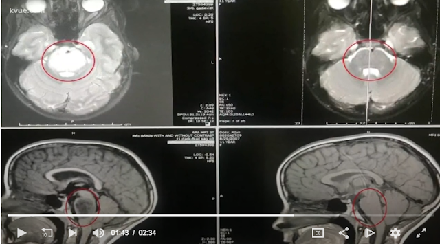
Disappearing pontine glioma


A little girl’s inoperable brain tumor is gone and doctors have no explanation.

Today, Roxli Doss is doing what she loves, and that’s horseback riding.

"She is just as active as she ever was,” said Scott Doss, Roxli’s father.

It's hard to imagine not too long ago doctors diagnosed the little girl with an inoperable brain tumor in June called diffuse intrinsic pontine glioma, or DIPG.

"It is very rare, but when we see it, it is a devastating disease,” Dr. Virginia Harrod with Dell Children’s Medical Center. “You have decreased ability to swallow, sometimes vision loss, decreased ability to talk, eventually difficulty with breathing."


Dr. Harrod said the now 11-year-old went through weeks of radiation, even though there is no cure. The family held a benefit for her in August, and the Buda community responded in a big way. At that point, all Gena and Scott Doss could do was pray for a miracle.

“And we got it,” said Gena.

“Praise God we did,” said Scott.

Now, they cry tears of joy.

"When I first saw Roxli's MRI scan, it was actually unbelievable,” said Dr. Harrod. “The tumor is undetectable on the MRI scan, which is really unusual.”

Doctors can't explain why the tumor disappeared.

"At Dell Children’s, Texas Children’s, at Dana-Farber, at John Hopkins, and MD Anderson, all agreed it was DIPG,” said Scott.

From no cure to no trace, the family said now they only have God to thank.

"Everyday we still say it,” said Gena. “It's kind of our family thing that God healed Roxli."

“We didn't know how long she would be healthy and, look at her, she's just doing awesome,” said Scott.

The parents said doctors double checked her scans just to confirm the results. Right now, they are watching her closely and she will continue to undergo treatments, such as immunotherapy, as a precaution.

https://www.kvue.com/article/news/local/from-no-cure-to-no-trace-central-texas-girls-inoperable-brain-tumor-disappears/269-623770908

https://www.foxnews.com/health/christmas-miracle-texas-girls-inoperable-brain-tumor-vanishes

Monday, December 17, 2018

Neuropsychological outcomes of children 1 year after pediatric cardiac arrest


Slomine BS, Silverstein FS, Christensen JR, Page K, Holubkov R, Dean JM, Moler FW. Neuropsychological Outcomes of Children 1 Year After Pediatric Cardiac Arrest: Secondary Analysis of 2 Randomized Clinical Trials. JAMA Neurol. 2018 Dec 1;75(12):1502-1510.

Abstract

IMPORTANCE:
Little is known about neuropsychological outcomes of children who survived pediatric cardiac arrest (CA).

OBJECTIVE:
To describe the neuropsychological outcomes of CA survivors enrolled in the Therapeutic Hypothermia After Pediatric Cardiac Arrest In-Hospital (THAPCA-IH) and Out-of-Hospital (THAPCA-OH) trials and compare the results with the primary outcome measure for these trials.

DESIGN, SETTING, AND PARTICIPANTS:
Secondary analysis of 222 CA survivors aged 1 to 18 years who received chest compressions for 2 minutes or more, remained comatose and required mechanical ventilation after return of circulation, and were enrolled in targeted temperature-management trials from 41 pediatric intensive care units. Data were collected from September 3, 2009, to February 3, 2016, and analyzed from March 10, 2017, to April 20, 2018.

MAIN OUTCOMES AND MEASURES:
The Vineland Adaptive Behavior Scales, Second Edition (VABS-II), a standardized measure of neurobehavioral functioning based on caregiver report (age-corrected mean [SD] scores = 100 [15]), was used to evaluate pre-CA functioning within 24 hours after enrollment; VABS-II<70 indicated significant developmental delays; VABS-II and neuropsychological testing were completed 1 year after CA. Neuropsychological testing included the Mullen Scales of Early Learning (Mullen) for children younger than 6 years and the Wechsler Abbreviated Scale of Intelligence (WASI) and neuropsychological measures of attention, memory, processing speed, and executive functioning for older children.

RESULTS:
Of 160 participants who completed neuropsychological testing, 96 (60.0%) were male; the median (interquartile range [IQR]) age was 2.5 years (1.3-6.1 years). Ninety-six (60.0%) were white, 41 (25.6%) were black, and 23 (14.4%) were of other/unknown race; 343 (21.2%) were Hispanic or Latino; 119 (74.4%) were non-Hispanic or Latino; and 7 (4.4%) were of unknown ethnicity. One hundred fourteen participants (71.2%) were classified as having favorable outcomes (VABS-II ≥70). Impairments (>2 SD below the mean for age) across neuropsychological measures ranged from 7% to 61%. Correlations between global cognitive and VABS-II scores were strong for younger children (Mullen, r = 0.69-0.87) but moderate for older children (r = 0.21-0.54 for the WASI). Of 111 children with favorable outcomes on VABS-II, 25.2% had global cognitive impairment and 30 of 35 older children (85.7%) had selective neuropsychological deficits.

CONCLUSIONS AND RELEVANCE:
In this prospectively evaluated cohort of pediatric CA survivors who were initially comatose, although 71.2% were classified as having favorable outcomes, significant neuropsychological deficits were identified in pediatric CA survivors who were classified as having favorable outcomes. The findings provide clinicians with a greater understanding of the spectrum of neuropsychological outcomes of pediatric CA survivors and the complex relationship between standardized caregiver-reported functional outcome measures incorporated in clinical trials and performance-based neuropsychological assessments.
_______________________________________________________________________ 

Researchers performed secondary analysis of cardiac arrest (CA) survivors enrolled in the Therapeutic Hypothermia After Pediatric Cardiac Arrest In-Hospital (THAPCA-IH) and Out-of-Hospital (THAPCA-OH) trials, to describe their neuropsychological outcomes. They also compare the results with the primary outcome measure for these trials. Although 71.2% of the survivors had favorable caregiver-rated outcomes 1 year later, the survivors displayed significant performance-based neuropsychological deficits across measures. 

Methods

Participants of this secondary analysis comprised 222 CA survivors, aged 1 to 18 years who received chest compressions for 2 minutes or more, remained comatose and required mechanical ventilation after return of circulation.

These participants were enrolled in targeted temperature-management trials from 41 pediatric intensive care units.

Collection of data was done from September 3, 2009, to February 3, 2016, and analysis was done from March 10, 2017, to April 20, 2018.

Pre-CA functioning within 24 hours after enrollment was evaluated using the Vineland Adaptive Behavior Scales, Second Edition (VABS-II), a standardized measure of neurobehavioral functioning based on caregiver report (age-corrected mean [SD] scores = 100 [15]); VABS-II<70 indicated significant developmental delays; completion of VABS-II and neuropsychological testing was done 1 year after CA.

The Mullen Scales of Early Learning (Mullen) was used for neuropsychological testing in children younger than 6 years; neuropsychological testing in older children was done determining the Wechsler Abbreviated Scale of Intelligence (WASI) and neuropsychological measures of attention, memory, processing speed, and executive functioning.

Results

Neuropsychological testing was completed by 160 participants [96 (60.0%) were male; the median (interquartile range [IQR]) age was 2.5 years (1.3-6.1 years)].

Of these, 96 (60.0%) were white, 41 (25.6%) were black, and 23 (14.4%) were of other/unknown race; 343 (21.2%) were Hispanic or Latino; 119 (74.4%) were non-Hispanic or Latino; and 7 (4.4%) were of unknown ethnicity.

Researchers classified 114 participants (71.2%) as having favorable outcomes (VABS-II ≥70).
The participants displayed impairments (>2 SD below the mean for age) ranging from 7% to 61% across neuropsychological measures.

Younger children displayed strong correlations (Mullen, r = 0.69-0.87) between global cognitive and VABS-II scores but older children showed moderate correlations (r = 0.21-0.54 for the WASI).
Global cognitive impairment was evident in 25.2% and selective neuropsychological deficits were noted in 30 of 35 older children (85.7%) among the 111 children with favorable outcomes on VABS-II.

https://www.mdlinx.com/journal-summaries/cardiology-child-development-critical-care-medicine/2018/12/12/7551094?spec=neurology